

Isthmin 1 is a secreted protein expressed in skin, mucosal tissues, and NK, NKT, and th17 cells
- PMID: 24956034
- PMCID: PMC4186767
- DOI: 10.1089/jir.2013.0137
Isthmin 1 is a secreted protein expressed in skin, mucosal tissues, and NK, NKT, and th17 cells
Abstract
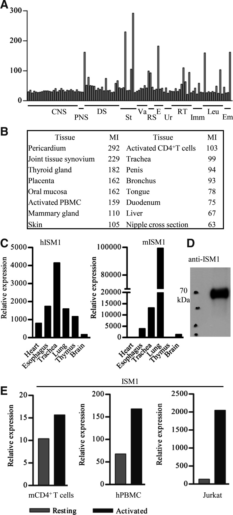
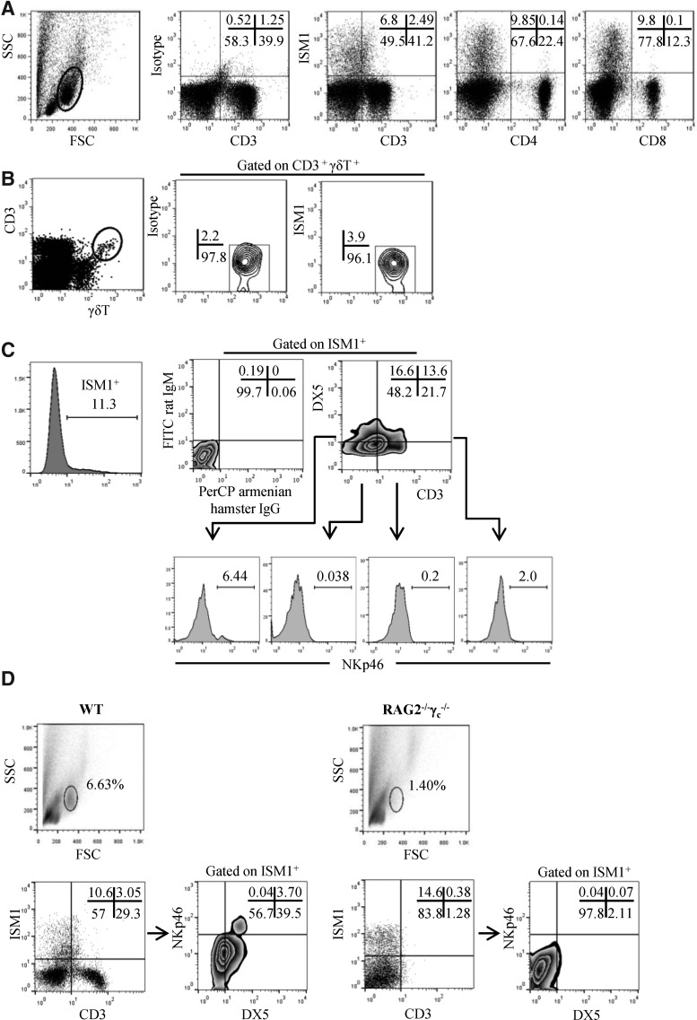
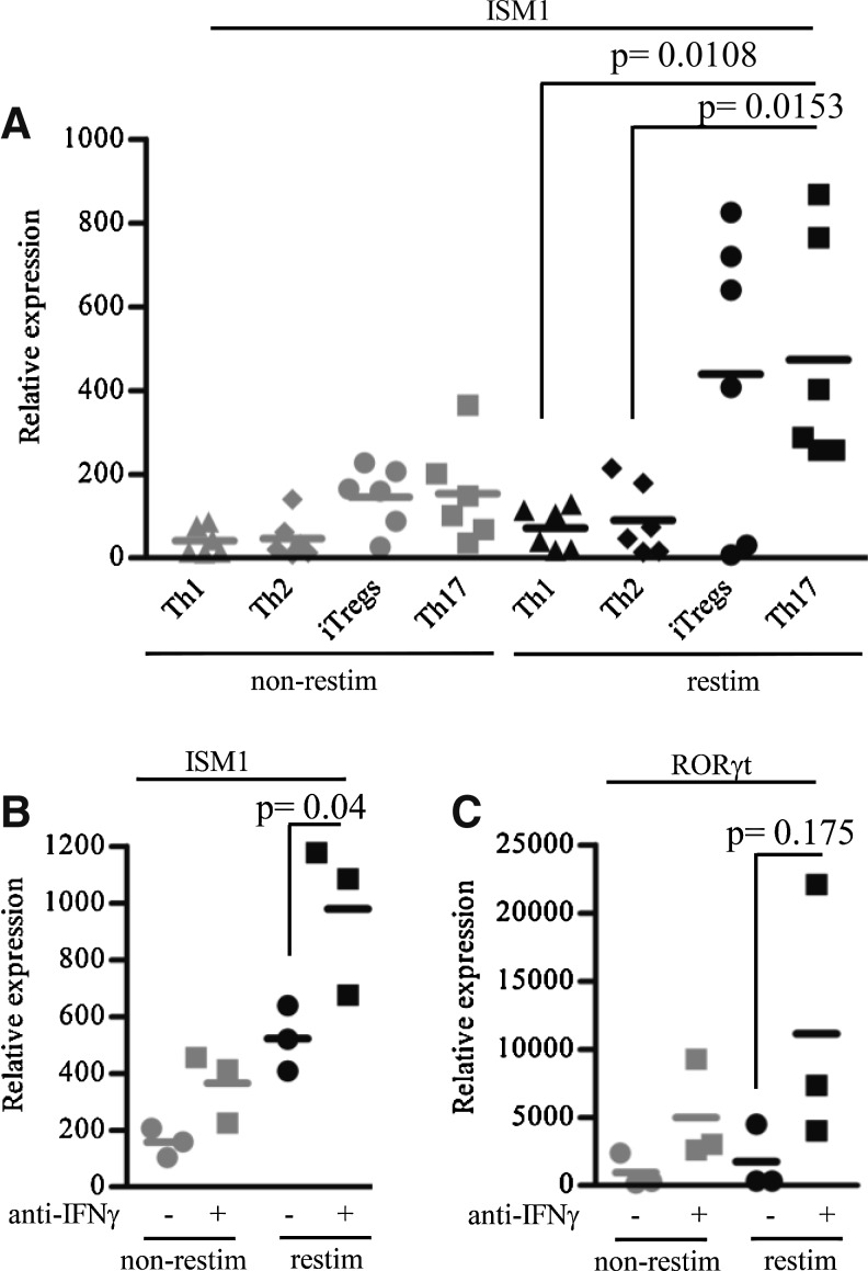
Using a comprehensive microarray database of human gene expression, we identified that in mammals, a secreted protein known as isthmin 1 (ISM1) is expressed in skin, mucosal tissues, and selected lymphocyte populations. ISM1 was originally identified in Xenopus brain during development, and it encodes a predicted ∼50-kDa protein containing a signal peptide, a thrombospondin domain, and an adhesion-associated domain. We confirmed the pattern of expression of ISM1 in both human and mouse tissues. ISM1 is expressed by DX5(+) lung lymphocytes that include NK and NKT-like cells, and is also expressed by some CD4(+) T cells upon activation but its expression increases significantly when CD4(+) T cells were polarized to the Th17 lineage in vitro. The presence of IFN-γ during CD4(+) T cell polarization inhibits ISM1 expression. Given that ISM1 has been reported to have anti-angiogenic properties, these observations suggest that ISM1 is a mediator of lymphocyte effector functions and may participate in both innate and acquired immune responses.
Figures
Similar articles
-
Regulation of N-glycosylation and secretion of Isthmin-1 by its C-mannosylation.Biochim Biophys Acta Gen Subj. 2021 Mar;1865(3):129840. doi: 10.1016/j.bbagen.2020.129840. Epub 2021 Jan 4. Biochim Biophys Acta Gen Subj. 2021. PMID: 33412225
-
DX5+NKT cells display phenotypical and functional differences between spleen and liver as well as NK1.1-Balb/c and NK1.1+ C57Bl/6 mice.BMC Immunol. 2011 Apr 29;12:26. doi: 10.1186/1471-2172-12-26. BMC Immunol. 2011. PMID: 21529347 Free PMC article.
-
Isthmin 1 is Expressed by Progenitor-Like Cells in the Lung: Phenotypical Analysis of Isthmin 1+ Hematopoietic Stem-Like Cells in Homeostasis and during Infection.J Immunol Res. 2022 Apr 1;2022:2909487. doi: 10.1155/2022/2909487. eCollection 2022. J Immunol Res. 2022. PMID: 35402623 Free PMC article.
-
Understanding the Regulatory Roles of Natural Killer T Cells in Rheumatoid Arthritis: T Helper Cell Differentiation Dependent or Independent?Scand J Immunol. 2016 Oct;84(4):197-203. doi: 10.1111/sji.12460. Scand J Immunol. 2016. PMID: 27384545 Review.
-
Mechanisms of innate lymphoid cell and natural killer T cell activation during mucosal inflammation.J Immunol Res. 2014;2014:546596. doi: 10.1155/2014/546596. Epub 2014 May 28. J Immunol Res. 2014. PMID: 24987710 Free PMC article. Review.
Cited by
-
METEORIN-LIKE is a cytokine associated with barrier tissues and alternatively activated macrophages.Clin Immunol. 2015 Feb;156(2):119-27. doi: 10.1016/j.clim.2014.11.006. Epub 2014 Dec 5. Clin Immunol. 2015. PMID: 25486603 Free PMC article.
-
Exploring the Functions of Mutant p53 through TP53 Knockout in HaCaT Keratinocytes.Curr Issues Mol Biol. 2024 Feb 8;46(2):1451-1466. doi: 10.3390/cimb46020094. Curr Issues Mol Biol. 2024. PMID: 38392212 Free PMC article.
-
Advances in research of biological functions of Isthmin-1.J Cell Commun Signal. 2023 Sep;17(3):507-521. doi: 10.1007/s12079-023-00732-3. Epub 2023 Mar 30. J Cell Commun Signal. 2023. PMID: 36995541 Free PMC article. Review.
-
Identification of IL-40, a Novel B Cell-Associated Cytokine.J Immunol. 2017 Nov 1;199(9):3326-3335. doi: 10.4049/jimmunol.1700534. Epub 2017 Oct 4. J Immunol. 2017. PMID: 28978694 Free PMC article.
-
The Association Between Serum Isthmin-1 and Disease Activity, Inflammation, and Autoantibody Status in Rheumatoid Arthritis.Diagnostics (Basel). 2025 May 23;15(11):1316. doi: 10.3390/diagnostics15111316. Diagnostics (Basel). 2025. PMID: 40506888 Free PMC article.
References
-
- Arase H, Saito T, Phillips JH, Lanier LL. 2001. Cutting edge: the mouse NK cell-associated antigen recognized by DX5 monoclonal antibody is CD49b (alpha 2 integrin, very late antigen-2). J Immunol 167(3):1141–1144 - PubMed
-
- Holt PG, Strickland DH, Wikstrom ME, Jahnsen FL. 2008. Regulation of immunological homeostasis in the respiratory tract. Nat Rev Immunol 8(2):142–152 - PubMed
-
- Ivanov II, McKenzie BS, Zhou L, Tadokoro CE, Lepelley A, Lafaille JJ, Cua DJ, Littman DR. 2006. The orphan nuclear receptor RORgammat directs the differentiation program of proinflammatory IL-17+ T helper cells. Cell 126(6):1121–1133 - PubMed
Publication types
MeSH terms
Substances
Grants and funding
LinkOut - more resources
Full Text Sources
Other Literature Sources
Research Materials
