. 2014 Aug 15;190(4):456-67.
doi: 10.1164/rccm.201403-0483OC.

Sex-dependent influence of endogenous estrogen in pulmonary hypertension

Affiliations

Sex-dependent influence of endogenous estrogen in pulmonary hypertension

Kirsty M Mair et al. Am J Respir Crit Care Med. .

Abstract

Rationale: The incidence of pulmonary arterial hypertension is greater in women, suggesting estrogens may play a role in the disease pathogenesis. Experimentally, in males, exogenously administered estrogen can protect against pulmonary hypertension (PH). However, in models that display female susceptibility, estrogens may play a causative role.

Objectives: To clarify the influence of endogenous estrogen and sex in PH and assess the therapeutic potential of a clinically available aromatase inhibitor.

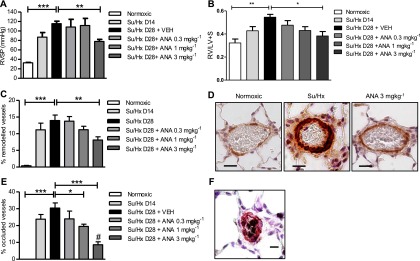
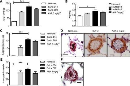
Methods: We interrogated the effect of reduced endogenous estrogen in males and females using the aromatase inhibitor, anastrozole, in two models of PH: the hypoxic mouse and Sugen 5416/hypoxic rat. We also determined the effects of sex on pulmonary expression of aromatase in these models and in lungs from patients with pulmonary arterial hypertension.

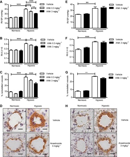
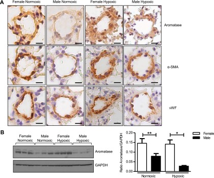
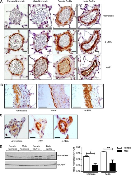
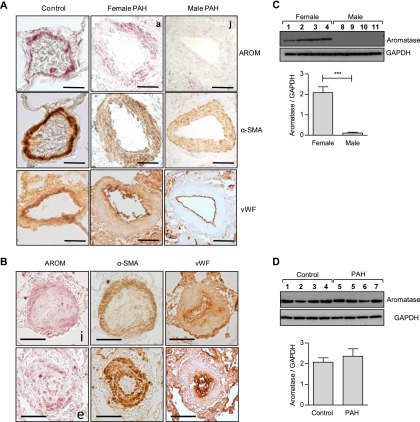
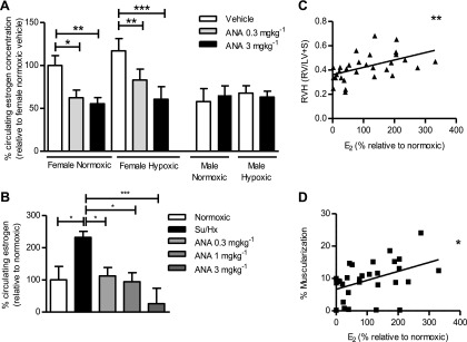
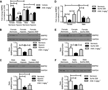
Measurements and main results: Anastrozole attenuated PH in both models studied, but only in females. To verify this effect was caused by reduced estrogenic activity we confirmed that in hypoxic mice inhibition of estrogen receptor α also has a therapeutic effect specifically in females. Female rodent lung displays increased aromatase and decreased bone morphogenetic protein receptor 2 and Id1 expression compared with male. Anastrozole treatment reversed the impaired bone morphogenetic protein receptor 2 pathway in females. Increased aromatase expression was also detected in female human pulmonary artery smooth muscle cells compared with male.

Conclusions: The unique phenotype of female pulmonary arteries facilitates the therapeutic effects of anastrozole in experimental PH confirming a role for endogenous estrogen in the disease pathogenesis in females and suggests aromatase inhibitors may have therapeutic potential.

Keywords: estrogen; pulmonary hypertension; sex.


Figures

Figure 1.
Figure 1.
Inhibition of aromatase attenuates chronic hypoxia-induced pulmonary hypertension (PH) in female mice. Female mice: Effects of the aromatase inhibitor, anastrozole (ANA), 0.3 mg ⋅ kg−1 ⋅ d−1 and 3 mg ⋅ kg−1 ⋅ d−1 for 14 days, on (A) right ventricular systolic pressure (RVSP) (n = 8–10 per group), (B) right ventricular hypertrophy (n = 8–10 per group) (as determined by RV/LV+S ratio), and (C) the % of remodeled pulmonary arteries in normoxic mice and hypoxic female mice with established PH (n = 6 per group). (D) Representative images of pulmonary arteries from normoxic and hypoxic female mice treated with or without anastrozole, 3 mg ⋅ kg−1 ⋅ d−1 (brown = α-smooth muscle actin; scale bar = 20 μm). Male mice: Effects of ANA, 3 mg ⋅ kg−1 ⋅ d−1 for 14 days, on (E) RVSP (n = 8–10 per group), (F) right ventricular hypertrophy (n = 8–10 per group) (as determined by RV/LV+S ratio), and (G) the % of remodeled pulmonary arteries in normoxic mice and hypoxic male mice with established PH (n = 6 per group). (H) Representative images of pulmonary arteries from normoxic and hypoxic male mice treated with or without anastrozole, 3 mg ⋅ kg−1 ⋅ d−1 (brown = α-smooth muscle actin; scale bar = 20 μm). Data displayed as mean ± SEM. **P < 0.01 and ***P < 0.001 as indicated, determined by one-way analysis of variance with Bonferroni post test. RV/LV+S = right ventricle/left ventricle + septum.
Figure 2.
Figure 2.
Inhibition of aromatase attenuates Su5416/hypoxia (Su/Hx)-induced pulmonary hypertension (PH) in female rats. (A) Right ventricular systolic pressure (RVSP) (n = 5–8 per group), (B) right ventricular hypertrophy (n = 5–8 per group) (as determined by RV/LV+S), and (C) the % remodeled pulmonary arteries (n = 5–8 per group) were assessed on Day 14 (D14) and Day 28 (D28) following administration of Su/Hx in female rats treated with or without anastrozole (ANA), 0.3 mg ⋅ kg−1 ⋅ d−1, 1 mg ⋅ kg−1 ⋅ d−1, or 3 mg ⋅ kg−1 ⋅ d−1, for 14 days in female rats (from D14–28). Representative images showing (D) α-smooth muscle actin (α-SMA) staining in pulmonary arteries from Su/Hx female rats treated with or without ANA (α-SMA = brown; scale bar = 20 μm). (E) The percentage of pulmonary arteries that are fully occluded in female rats treated with or without ANA (n = 5–8) and (F) representative image of an occluded pulmonary artery (α-SMA = pink, von Willebrand factor = black; scale bar = 20 μm). Data displayed as mean ± SEM. *P < 0.05, **P < 0.01, and ***P < 0.001 as indicated, #P < 0.01 versus D14 Su/Hx as determined by one-way analysis of variance with Dunnett post test.
Figure 3.
Figure 3.
Inhibition of aromatase does not attenuate Su5416/hypoxia (Su/Hx)-induced pulmonary hypertension (PH) in male rats. (A) Right ventricular systolic pressure (RVSP) (n = 5–8 per group), (B) right ventricular hypertrophy (n = 5–8 per group), and (C) the % remodeled pulmonary arteries (n = 5–8 per group) were assessed on Day 14 (D14) and Day 28 (D28) following administration of Su/Hx in male rats treated with or without anastrozole (ANA), 3 mg ⋅ kg−1 ⋅ d−1 (from D14 to D28). Representative images showing (D) α-smooth muscle actin (α-SMA) staining in pulmonary arteries from SU/Hx male rats treated with or without anastrozole (α-SMA = brown; scale bar = 20 μm). (E) The percentage of pulmonary arteries that are fully occluded (n = 5–8 per group) in male rats treated with or without anastrozole, 3 mg ⋅ kg−1 ⋅ d−1, and (F) representative image of an occluded pulmonary artery (α-SMA = pink; von Willebrand factor = black ; scale bar = 10 μm). Data displayed as mean ± SEM. *P < 0.05 and ***P < 0.001 as indicated, determined by one-way analysis of variance with Dunnett post test.
Figure 4.
Figure 4.
Effect of chronic hypoxia on aromatase expression in mouse pulmonary artery and whole lung. Representative images showing (A) aromatase immunolocalization in pulmonary arteries (scale bar = 20 μm) with 3 μm consecutive sections showing α-smooth muscle actin (α-SMA) and von Willebrand factor (vWF) (representative of n = 4 per group, brown staining). For IgG control see Figure E5. (B) Representative immunoblot and quantification of aromatase protein expression in whole lung from normoxic and hypoxic, female and male mice (n = 5–6 per group). Data displayed as mean ± SEM. *P < 0.05 and **P < 0.01 as indicated, determined by one-way analysis of variance with Bonferroni post test. GAPDH = glyceraldehyde phosphate dehydrogenase.
Figure 5.
Figure 5.
Effect of Su5416/hypoxia (Su/Hx) on aromatase expression in rat pulmonary artery and whole lung. Representative images showing (A) aromatase immunolocalization in pulmonary arteries (scale bar = 20 μm) with 3 μm consecutive sections showing α-smooth muscle actin (α-SMA) and von Willebrand factor (vWF) (representative of n = 4 per group, brown staining) (for IgG control see Figure E5). (B) Representative image showing the absence of aromatase immunolocalization in the endothelial layer of rat pulmonary artery (scale bar = 50 μm [×400 magnification]) with 3 μm consecutive sections showing α-SMA and vWF (brown staining). (C) Representative image showing aromatase immunolocalization in small occlusive vascular lesions from SuHx rat (scale bar = 20 μm) with 3 μm consecutive sections showing α-SMA and vWF (brown staining). (D) Representative immunoblot and quantification of aromatase protein expression in whole lung from normoxic and hypoxic, female and male rats (n = 5–6 per group). Data displayed as mean ± SEM. *P < 0.05 and **P < 0.01 as indicated, determined by one-way analysis of variance with Bonferroni post test. GAPDH = glyceraldehyde phosphate dehydrogenase.
Figure 6.
Figure 6.
Aromatase (AROM) expression in human pulmonary arterial hypertension (PAH). (A) Representative images showing AROM immunolocalization in control and female and male patients with PAH. (B) Representative images showing examples of AROM immunolocalization in vascular lesions from patients with PAH (AROM = pink; α-smooth muscle actin [α-SMA] and von Willebrand factor [vWF] = brown; scale bar = 100 μm; for IgG control see Figure E5). AROM protein expression was also assessed by immunoblotting using human pulmonary artery smooth muscle cells. (C) Representative immunoblots and graph showing quantification comparing AROM expression in human pulmonary artery smooth muscle cells isolated from male and female control subjects (n = 4 samples per group) and (D) female control and female patients with PAH (n = 4 samples per group). Data displayed as mean ± SEM. ***P < 0.001 as indicated, determined by two-tailed, unpaired t test. 1–11, a, e, i, and j correspond to patient information on human tissues and cells referred to in Table E3. GAPDH = glyceraldehyde phosphate dehydrogenase.
Figure 7.
Figure 7.
Effect of aromatase inhibition on circulating estradiol (E2) levels in models of pulmonary hypertension. (A) Circulating plasma E2 levels in normoxic and hypoxic female and male mice treated with or without anastrozole (ANA) for 14 days (n = 5 per group). Data displayed as mean ± SEM. *P < 0.05, **P < 0.01, and ***P < 0.001 as indicated, determined by one-way analysis of variance with Bonferroni post test. (B) Circulating E2 levels in female Su/Hx rats treated with or without 0.3 mg ⋅ kg−1 ⋅ d−1, 1 mg ⋅ kg−1 ⋅ d−1, or 3 mg ⋅ kg−1 ⋅ d−1 ANA (n = 4–5 per group). Data displayed as mean ± SEM. *P < 0.05 and ***P < 0.001 as indicated, determined by one-way analysis of variance with Dunnett post test. Plasma E2 levels from female Su/Hx rats were used to determine if there was any correlation with disease severity using Pearson coefficient. Significant correlation between plasma E2 levels and (C) right ventricular hypertrophy (RVH) (as determined by RV/LV+S) (n = 30), and (D) the percentage of muscularized pulmonary arteries (n = 30). *P < 0.05 and **P < 0.01 as indicated. All E2 concentrations are expressed as a percentage relative to normoxic set at 100%.
Figure 8.
Figure 8.
Effects of aromatase inhibition on hypoxia and Su/Hx–induced changes in bone morphogenetic protein receptor 2 (BMPR2) expression. (A–C) Hypoxic mice. (A) Relative gene expression levels of BMPR2 in male and female normoxic and hypoxic mouse lung treated with or without anastrozole (ANA), 3 mg ⋅ kg−1 ⋅ d−1 (n = 6 per group). Representative immunoblot and quantification showing effects of ANA, 3 mg ⋅ kg−1 ⋅ d−1, on BMPR2 protein expression in (B) female and (C) male normoxic and hypoxic mouse lung (n = 6 per group). (D–F) Su/Hx rats. (D) Relative gene expression levels of BMPR2 in female and male normoxic and Su/Hx rat lung treated with or without ANA, 3 mg ⋅ kg−1 ⋅ d−1 for 14 days (n = 5–6 per group). Representative immunoblot and quantification showing effects of ANA, 3 mg ⋅ kg−1 ⋅ d−1, on BMPR2 protein expression in (E) female and (F) male lung from normoxic and Su/Hx rats treated with or without ANA, 3 mg ⋅ kg−1 ⋅ d−1 for 14 days (n = 5–6 per group). Gene expression levels are normalized to β2-microglobulin (β2M). Data displayed as mean ± SEM. *P < 0.05, **P < 0.01, and ***P < 0.001 as indicated, determined by one-way analysis of variance with Bonferroni post test. GAPDH = glyceraldehyde phosphate dehydrogenase.

References

    1. Shapiro S, Traiger GL, Turner M, McGoon MD, Wason P, Barst RJ. Sex differences in the diagnosis, treatment, and outcome of patients with pulmonary arterial hypertension enrolled in the registry to evaluate early and long-term pulmonary arterial hypertension disease management. Chest. 2012;141:363–373. - PubMed
    1. McGoon MD, Benza RL, Escribano-Subias P, Jiang X, Miller DP, Peacock AJ, Pepke-Zaba J, Pulido T, Rich S, Rosenkranz S, et al. Pulmonary arterial hypertension: epidemiology and registries. J Am Coll Cardiol. 2013;62(Suppl. 25):D51–D59. - PubMed
    1. Rabinovitch M. Molecular pathogenesis of pulmonary arterial hypertension. J Clin Invest. 2012;122:4306–4313. - PMC - PubMed
    1. Lahm T, Albrecht M, Fisher AJ, Selej M, Patel NG, Brown JA, Justice MJ, Brown MB, Van Demark M, Trulock KM, et al. 17β-Estradiol attenuates hypoxic pulmonary hypertension via estrogen receptor-mediated effects. Am J Respir Crit Care Med. 2012;185:965–980. - PMC - PubMed
    1. Umar S, Iorga A, Matori H, Nadadur RD, Li J, Maltese F, van der Laarse A, Eghbali M. Estrogen rescues preexisting severe pulmonary hypertension in rats. Am J Respir Crit Care Med. 2011;184:715–723. - PMC - PubMed

Publication types

MeSH terms