

Is macroporosity absolutely required for preliminary in vitro bone biomaterial study? A comparison between porous materials and flat materials
- PMID: 24956447
- PMCID: PMC4030915
- DOI: 10.3390/jfb2040308
Is macroporosity absolutely required for preliminary in vitro bone biomaterial study? A comparison between porous materials and flat materials
Abstract
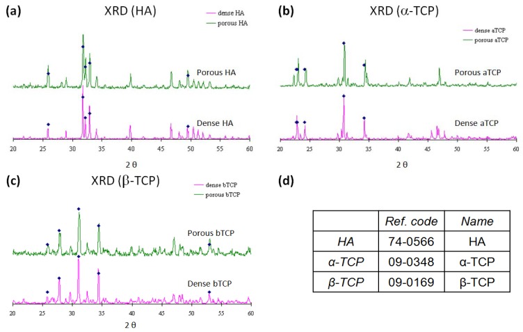
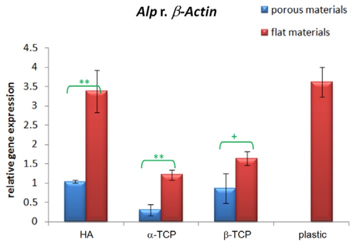
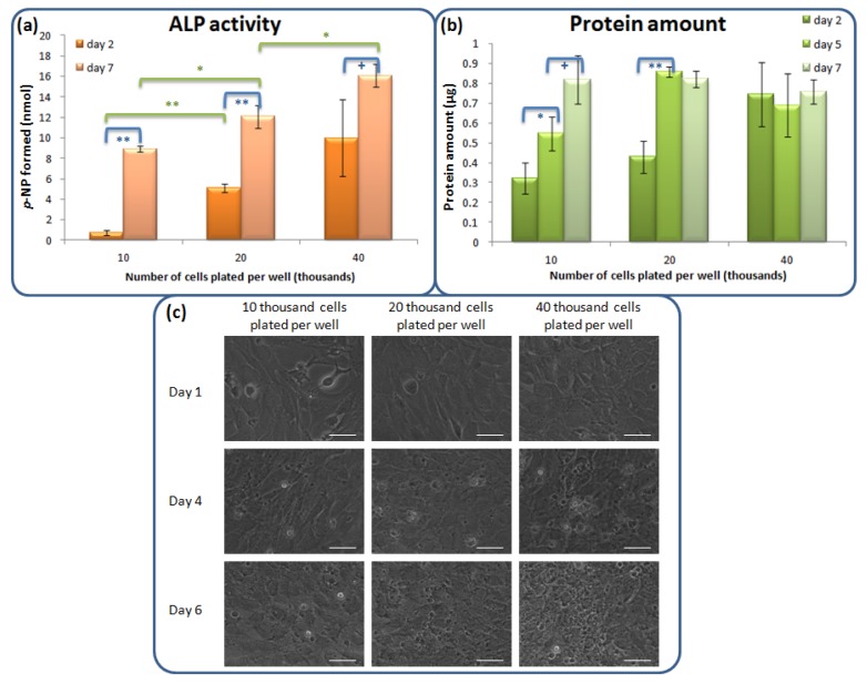
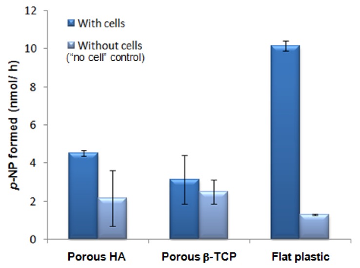
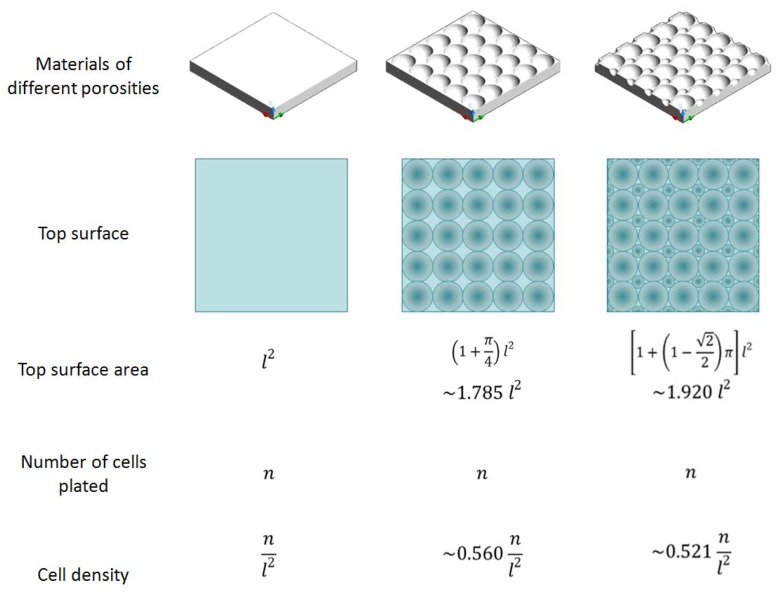
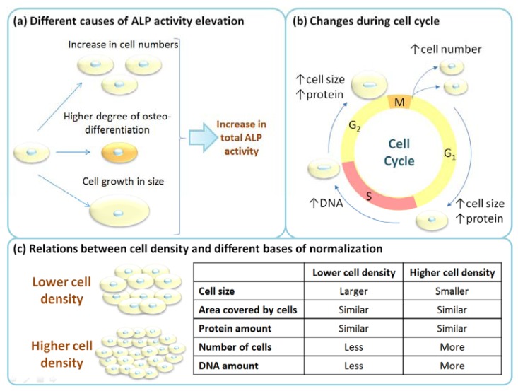
Porous materials are highly preferred for bone tissue engineering due to space for blood vessel ingrowth, but this may introduce extra experimental variations because of the difficulty in precise control of porosity. In order to decide whether it is absolutely necessary to use porous materials in in vitro comparative osteogenesis study of materials with different chemistries, we carried out osteoinductivity study using C3H/10T1/2 cells, pluripotent mesenchymal stem cells (MSCs), on seven material types: hydroxyapatite (HA), α-tricalcium phosphate (α-TCP) and b-tricalcium phosphate (β-TCP) in both porous and dense forms and tissue culture plastic. For all materials under test, dense materials give higher alkaline phosphatase gene (Alp) expression compared with porous materials. In addition, the cell density effects on the 10T1/2 cells were assessed through alkaline phosphatase protein (ALP) enzymatic assay. The ALP expression was higher for higher initial cell plating density and this explains the greater osteoinductivity of dense materials compared with porous materials for in vitro study as porous materials would have higher surface area. On the other hand, the same trend of Alp mRNA level (HA > β-TCP > α-TCP) was observed for both porous and dense materials, validating the use of dense flat materials for comparative study of materials with different chemistries for more reliable comparison when well-defined porous materials are not available. The avoidance of porosity variation would probably facilitate more reproducible results. This study does not suggest porosity is not required for experiments related to bone regeneration application, but emphasizes that there is often a tradeoff between higher clinical relevance, and less variation in a less complex set up, which facilitates a statistically significant conclusion. Technically, we also show that the base of normalization for ALP activity may influence the conclusion and there may be ALP activity from serum, necessitating the inclusion of "no cell" control in ALP activity assay with materials. These explain the opposite conclusions drawn by different groups on the effect of porosity.
Figures


Similar articles
-
Hollow calcium phosphate microcarriers for bone regeneration: in vitro osteoproduction and ex vivo mechanical assessment.Biomed Mater Eng. 2007;17(5):277-89. Biomed Mater Eng. 2007. PMID: 17851170
-
Ectopic osteoinduction and early degradation of recombinant human bone morphogenetic protein-2-loaded porous beta-tricalcium phosphate in mice.Biomaterials. 2005 Jul;26(20):4265-71. doi: 10.1016/j.biomaterials.2004.10.035. Biomaterials. 2005. PMID: 15683650
-
Preparation and in vitro evaluation of mesoporous hydroxyapatite coated β-TCP porous scaffolds.Mater Sci Eng C Mater Biol Appl. 2013 Dec 1;33(8):5001-7. doi: 10.1016/j.msec.2013.08.027. Epub 2013 Aug 31. Mater Sci Eng C Mater Biol Appl. 2013. PMID: 24094217
-
Effects of porous beta-tricalcium phosphate-based ceramics used as an E. coli-derived rhBMP-2 carrier for bone regeneration.J Mater Sci Mater Med. 2013 Sep;24(9):2117-27. doi: 10.1007/s10856-013-4967-5. Epub 2013 Jun 1. J Mater Sci Mater Med. 2013. PMID: 23728522
-
Porosity and pore size of beta-tricalcium phosphate scaffold can influence protein production and osteogenic differentiation of human mesenchymal stem cells: an in vitro and in vivo study.Acta Biomater. 2008 Nov;4(6):1904-15. doi: 10.1016/j.actbio.2008.05.017. Epub 2008 Jun 11. Acta Biomater. 2008. PMID: 18571999
Cited by
-
Natural bone-mimicking nanopore-incorporated hydroxyapatite scaffolds for enhanced bone tissue regeneration.Biomater Res. 2022 Feb 25;26(1):7. doi: 10.1186/s40824-022-00253-x. Biomater Res. 2022. PMID: 35216625 Free PMC article.
-
Fabrication of 3D-Printed Scaffolds with Multiscale Porosity.ACS Omega. 2024 Jun 28;9(27):29186-29204. doi: 10.1021/acsomega.3c09035. eCollection 2024 Jul 9. ACS Omega. 2024. PMID: 39005818 Free PMC article.
-
Tunable Properties via Composition Modulations of Poly(vinyl alcohol)/Xanthan Gum/Oxalic Acid Hydrogels.Materials (Basel). 2022 Apr 4;15(7):2657. doi: 10.3390/ma15072657. Materials (Basel). 2022. PMID: 35407989 Free PMC article.
-
Ceramic Biomaterial Pores Stereology Analysis by the Use of Microtomography.Materials (Basel). 2021 Apr 25;14(9):2207. doi: 10.3390/ma14092207. Materials (Basel). 2021. PMID: 33923089 Free PMC article.
References
-
- Karageorgiou V., Kaplan D. Porosity of 3D biomaterial scaffolds and osteogenesis. Biomaterials. 2005;26:5474–5491. - PubMed
-
- Peng Q., Jiang F., Huang P., Zhou S., Weng J., Bao C., Zhang C., Yu H. A novel porous bioceramics scaffold by accumulating hydroxyapatite spherules for large bone tissue engineering in vivo. I. Preparation and characterization of scaffold. J. Biomed. Mater. Res. Part A. 2010;93:920–929. - PubMed
-
- Yun H.-S., Kim S.-E., Park E.K. Bioactive glass-poly (ε-caprolactone) composite scaffolds with 3 dimensionally hierarchical pore networks. Mater. Sci. Eng. C. 2011;31:198–205.
-
- Tang P.-F., Li G., Wang J.-F., Zheng Q.-J., Wang Y. Development, characterization, and validation of porous carbonated hydroxyapatite bone cement. J. Biomed. Mater. Res. Part B. 2009;90:886–893. - PubMed
LinkOut - more resources
Full Text Sources
