

Biocompatible polymer/quantum dots hybrid materials: current status and future developments
- PMID: 24956449
- PMCID: PMC4030917
- DOI: 10.3390/jfb2040355
Biocompatible polymer/quantum dots hybrid materials: current status and future developments
Abstract
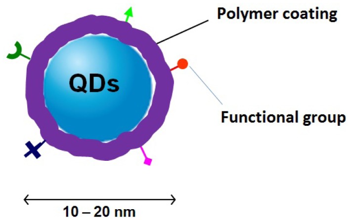
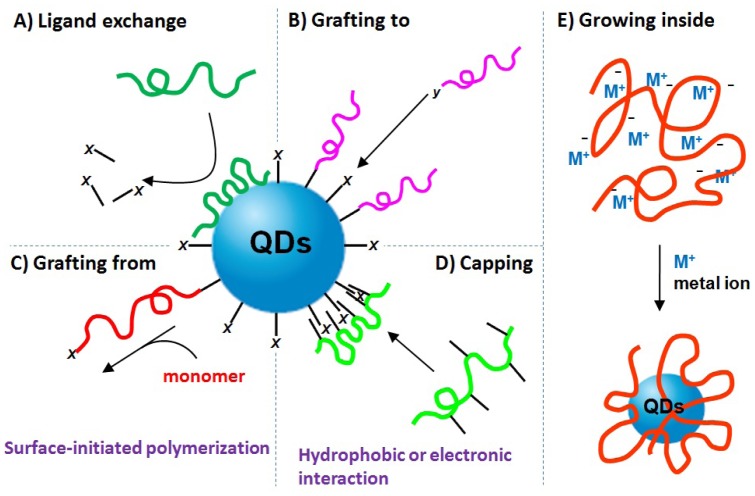
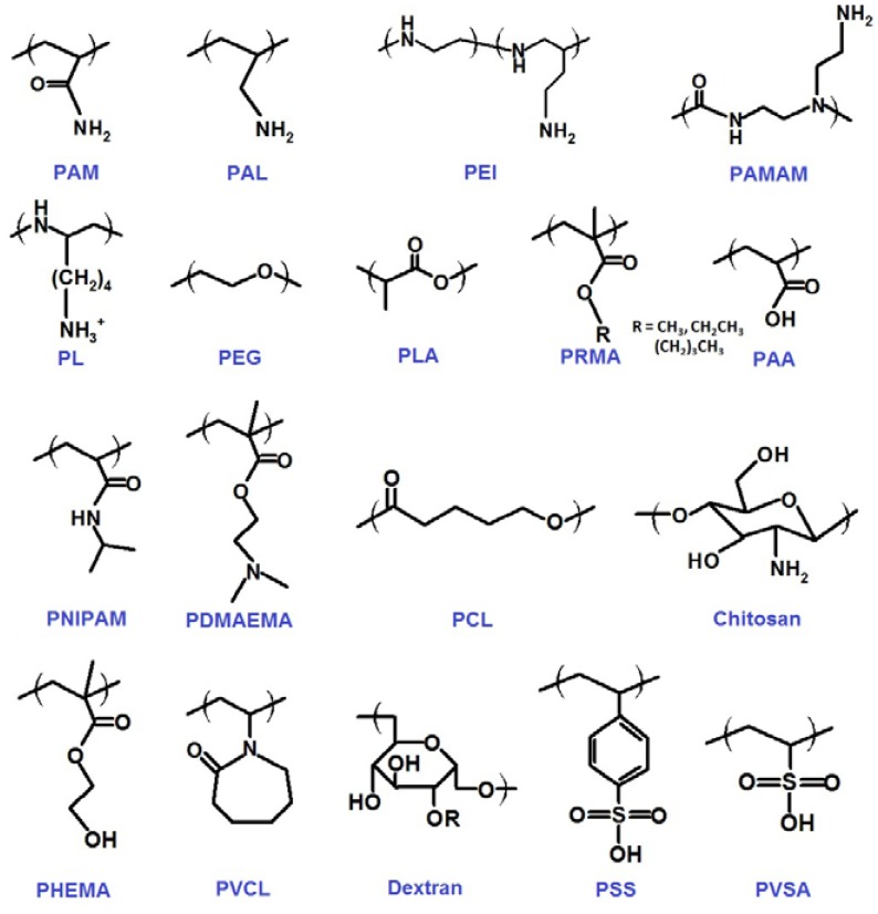
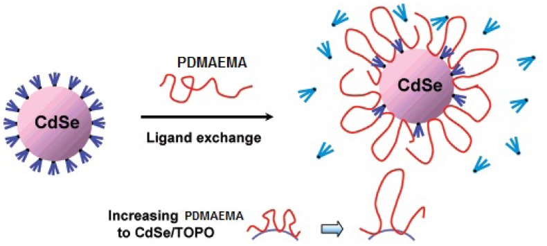
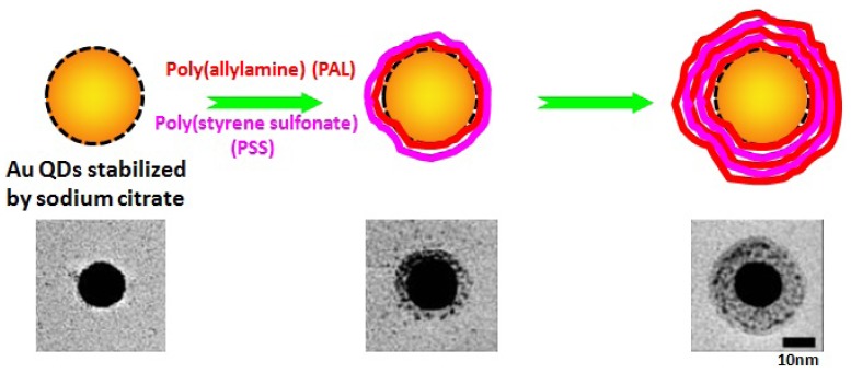
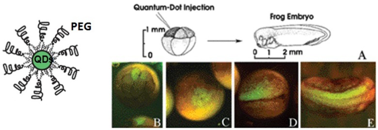
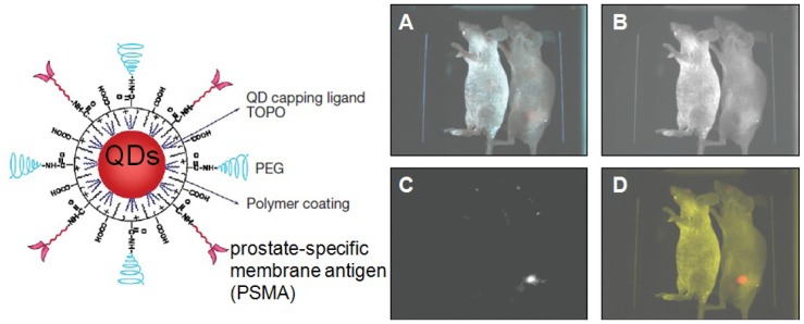
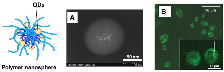
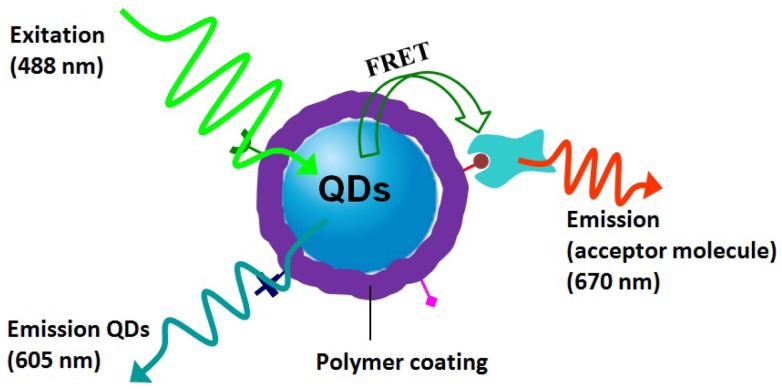
Quantum dots (QDs) are nanometer-sized semiconductor particles with tunable fluorescent optical property that can be adjusted by their chemical composition, size, or shape. In the past 10 years, they have been demonstrated as a powerful fluorescence tool for biological and biomedical applications, such as diagnostics, biosensing and biolabeling. QDs with high fluorescence quantum yield and optical stability are usually synthesized in organic solvents. In aqueous solution, however, their metallic toxicity, non-dissolubility and photo-luminescence instability prevent the direct utility of QDs in biological media. Polymers are widely used to cover and coat QDs for fabricating biocompatible QDs. Such hybrid materials can provide solubility and robust colloidal and optical stability in water. At the same time, polymers can carry ionic or reactive functional groups for incorporation into the end-use application of QDs, such as receptor targeting and cell attachment. This review provides an overview of the recent development of methods for generating biocompatible polymer/QDs hybrid materials with desirable properties. Polymers with different architectures, such as homo- and co-polymer, hyperbranched polymer, and polymeric nanogel, have been used to anchor and protect QDs. The resulted biocompatible polymer/QDs hybrid materials show successful applications in the fields of bioimaging and biosensing. While considerable progress has been made in the design of biocompatible polymer/QDs materials, the research challenges and future developments in this area should affect the technologies of biomaterials and biosensors and result in even better biocompatible polymer/QDs hybrid materials.
Figures
Similar articles
-
Polymer coating of quantum dots--a powerful tool toward diagnostics and sensorics.Eur J Pharm Biopharm. 2008 Jan;68(1):138-52. doi: 10.1016/j.ejpb.2007.05.013. Epub 2007 Jun 15. Eur J Pharm Biopharm. 2008. PMID: 17689938 Review.
-
Colloidal synthesis of tunably luminescent AgInS-based/ZnS core/shell quantum dots as biocompatible nano-probe for high-contrast fluorescence bioimaging.Mater Sci Eng C Mater Biol Appl. 2020 Jun;111:110807. doi: 10.1016/j.msec.2020.110807. Epub 2020 Mar 3. Mater Sci Eng C Mater Biol Appl. 2020. PMID: 32279757
-
Fluorescent cadmium telluride quantum dots embedded chitosan nanoparticles: a stable, biocompatible preparation for bio-imaging.J Biomater Sci Polym Ed. 2015;26(1):42-56. doi: 10.1080/09205063.2014.982240. Epub 2014 Nov 20. J Biomater Sci Polym Ed. 2015. PMID: 25410797
-
Biosynthesized Quantum Dots as Improved Biocompatible Tools for Biomedical Applications.Curr Med Chem. 2021;28(3):496-513. doi: 10.2174/0929867327666200102122737. Curr Med Chem. 2021. PMID: 31894739 Review.
-
Bioconjugated quantum dots as fluorescent probes for bioanalytical applications.Anal Bioanal Chem. 2010 Jan;396(1):229-40. doi: 10.1007/s00216-009-3033-0. Epub 2009 Aug 28. Anal Bioanal Chem. 2010. PMID: 19714321 Review.
Cited by
-
Poly(ferulic acid-co-tyrosine): Effect of the Regiochemistry on the Photophysical and Physical Properties en Route to Biomedical Applications.Macromolecules. 2014 Oct 28;47(20):7109-7117. doi: 10.1021/ma5015534. Epub 2014 Oct 14. Macromolecules. 2014. PMID: 25364040 Free PMC article.
-
Recent advances in rapid pathogen detection method based on biosensors.Eur J Clin Microbiol Infect Dis. 2018 Jun;37(6):1021-1037. doi: 10.1007/s10096-018-3230-x. Epub 2018 Mar 22. Eur J Clin Microbiol Infect Dis. 2018. PMID: 29569045 Review.
-
Hybrid Nanomat: Copolymer Template CdSe Quantum Dots In Situ Stabilized and Immobilized within Nanofiber Matrix.Nanomaterials (Basel). 2023 Feb 5;13(4):630. doi: 10.3390/nano13040630. Nanomaterials (Basel). 2023. PMID: 36838998 Free PMC article.
-
Extremely high brightness from polymer-encapsulated quantum dots for two-photon cellular and deep-tissue imaging.Sci Rep. 2015 Apr 24;5:9908. doi: 10.1038/srep09908. Sci Rep. 2015. PMID: 25909393 Free PMC article.
-
Optical and Physicochemical Characterizations of a Cellulosic/CdSe-QDs@S-DAB5 Film.Nanomaterials (Basel). 2022 Jan 29;12(3):484. doi: 10.3390/nano12030484. Nanomaterials (Basel). 2022. PMID: 35159829 Free PMC article.
References
-
- Nirmal M., Brus L. Luminescence photophysics in semiconductor nanocrystals. Acc. Chem. Res. 1999;32:407–414.
-
- Tomczak N., Jańczewski D., Han M., Vancso G.J. Designer polymer-quantum dots architectures. Prog. Polym. Sci. 2009;34:393–430.
-
- Hezinger A.F.E., Teβmar J., Göpferich A. Polymer coating of quantum dots—A powerful tool toward diagnostics and sensorics. E. J. Pharm. Biopharm. 2008;68:138–152. - PubMed
LinkOut - more resources
Full Text Sources
Other Literature Sources
Miscellaneous
