

Deficiency of formyl peptide receptor 1 and 2 is associated with increased inflammation and enhanced liver injury after LPS-stimulation
- PMID: 24956481
- PMCID: PMC4067326
- DOI: 10.1371/journal.pone.0100522
Deficiency of formyl peptide receptor 1 and 2 is associated with increased inflammation and enhanced liver injury after LPS-stimulation
Abstract
Introduction: Formyl peptide-receptor 1 and 2 (FPR1 and FPR2) in mice were identified as receptors with contrary affinity for the PAMP fMLF. Formyl-methionyl-leucyl-phenylalanine is either part of the bacterial membrane and is secreted by the mitochondria of eukaryotic ceslls during apoptosis. Furthermore FPR1 and 2 are described as highly relevant factors for the chemotaxis of immune cells. Their role during the acute liver injury has not been investigated yet.
Materials and methods: Constitutive knockout mice for FPR1 (mFPR1-/-), FPR2 (mFPR2-/-) and wild type (WT) mice were challenged with LPS i.p. for 3 h and 6 h. Liver and serum were sampled for further analysis.
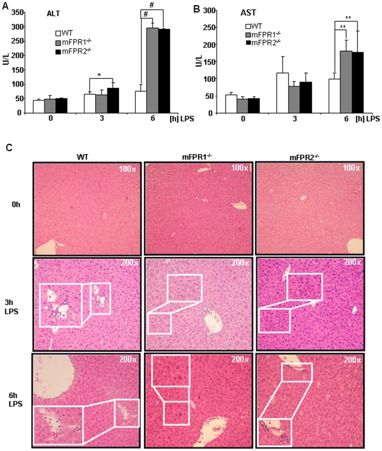
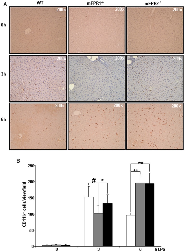
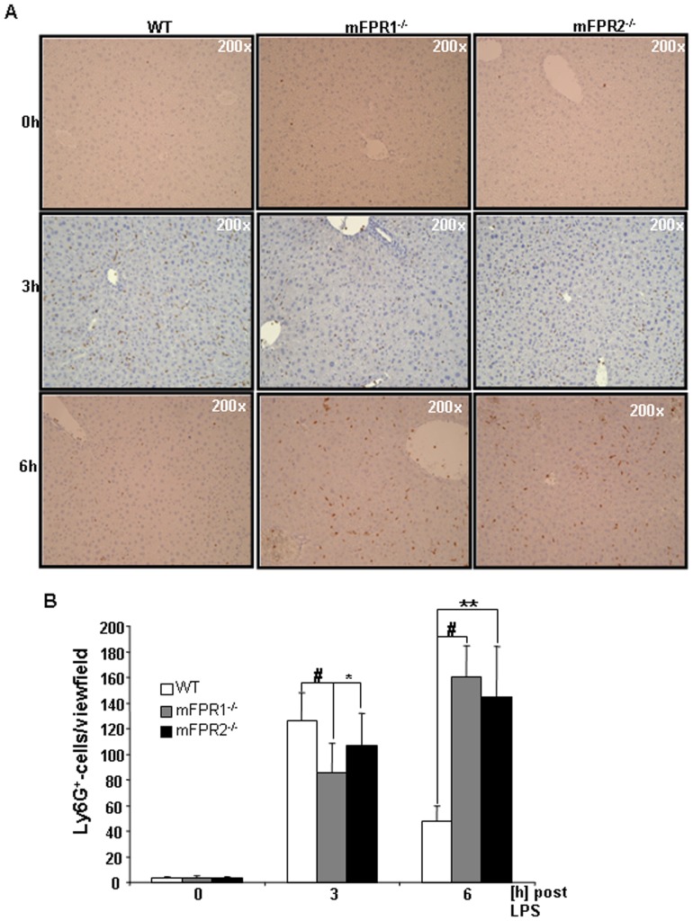
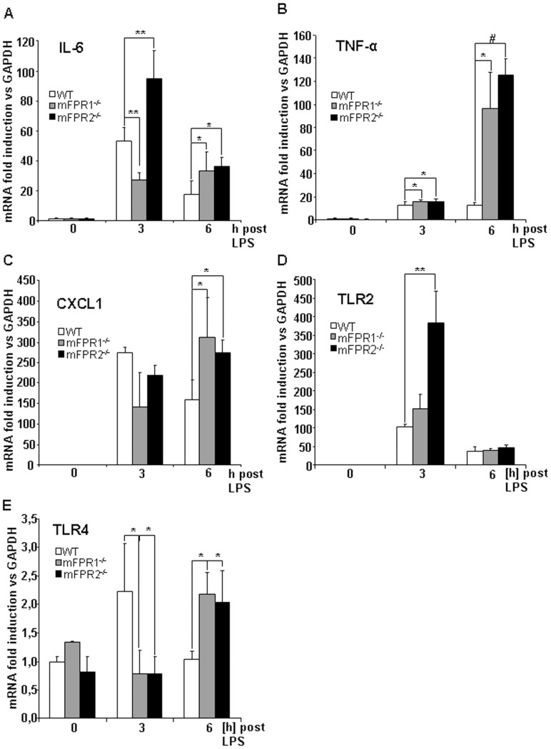
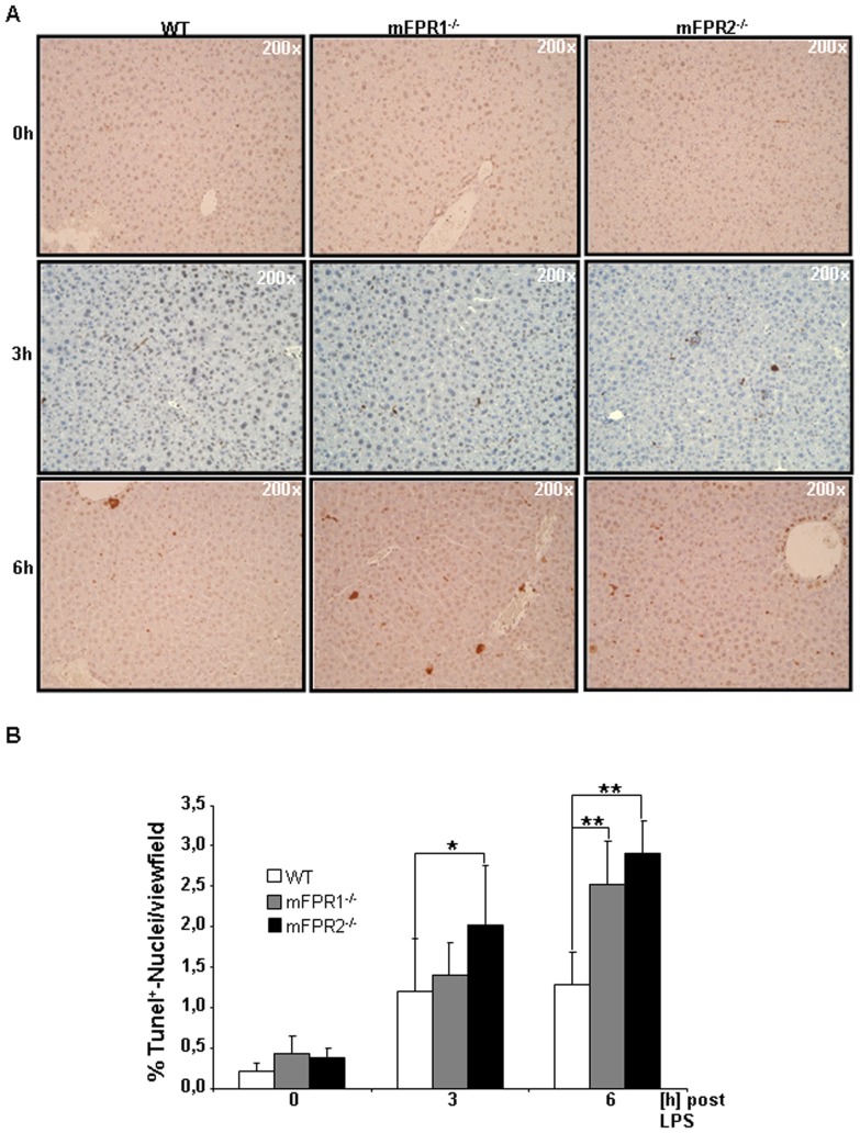
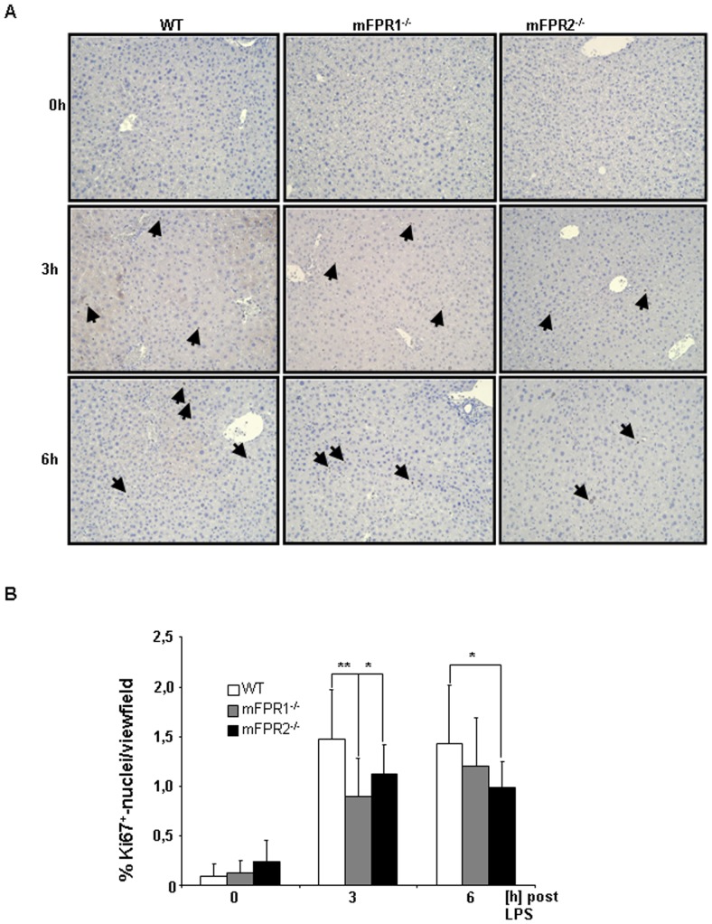
Results: Liver transaminases were elevated in all mice 3 h and 6 h post LPS stimulation. Gene expression analysis displayed a reduced expression of the pro-inflammatory cytokines IL-6 and CXCL1 after 3 h in the mFPR1-/- compared to wild type and mFPR2-/- mice. After 6 h, IL-6, TNF-α and CXCL1 were significantly higher in mice lacking mFPR1 or 2. Consistent to these findings the numbers of CD11b+ and Ly6G+ immune cells were altered in the livers. The analysis of TLR2 and TLR4 revealed time and genotype specific changes in theirs gene expression. Additionally, the liver in mFPR1- and mFPR2-deficient mice seem to be more susceptible to apoptosis by showing a significant higher number of TUNEL+-cells in the liver than WT-mice and displayed less Ki67-positive nuclei in the liver.
Conclusion: The results suggest a prominent role of FPRs in the regulation of the hepatic inflammatory response after LPS induced liver injury. Deletion of mFPR1 or mFPR2 leads to deregulation of the inflammatory response compared to WT mice, associated with more severe liver injury represented by higher levels of transaminases, apoptotic cells and a reduced regenerative capacity.
Conflict of interest statement
Figures
Similar articles
-
RhoA/ROCK downregulates FPR2-mediated NADPH oxidase activation in mouse bone marrow granulocytes.Cell Signal. 2014 Oct;26(10):2138-46. doi: 10.1016/j.cellsig.2014.05.017. Epub 2014 May 29. Cell Signal. 2014. PMID: 24880063
-
The formyl peptide fMLF primes platelet activation and augments thrombus formation.J Thromb Haemost. 2019 Jul;17(7):1120-1133. doi: 10.1111/jth.14466. Epub 2019 May 24. J Thromb Haemost. 2019. PMID: 31033193 Free PMC article.
-
Lipopolysaccharide-induced liver apoptosis is increased in interleukin-10 knockout mice.Biochim Biophys Acta. 2006 Apr;1762(4):468-77. doi: 10.1016/j.bbadis.2005.12.012. Epub 2006 Feb 13. Biochim Biophys Acta. 2006. PMID: 16497487
-
The Contribution of Chemoattractant GPCRs, Formylpeptide Receptors, to Inflammation and Cancer.Front Endocrinol (Lausanne). 2020 Jan 24;11:17. doi: 10.3389/fendo.2020.00017. eCollection 2020. Front Endocrinol (Lausanne). 2020. PMID: 32038501 Free PMC article. Review.
-
Formyl peptide receptor 2 and heart disease.Semin Immunol. 2022 Jan;59:101602. doi: 10.1016/j.smim.2022.101602. Epub 2022 Mar 8. Semin Immunol. 2022. PMID: 35277300 Review.
Cited by
-
Formyl peptide receptor 2 determines sex-specific differences in the progression of nonalcoholic fatty liver disease and steatohepatitis.Nat Commun. 2022 Jan 31;13(1):578. doi: 10.1038/s41467-022-28138-6. Nat Commun. 2022. PMID: 35102146 Free PMC article.
-
Fpr2/CXCL1/2 Controls Rapid Neutrophil Infiltration to Inhibit Streptococcus agalactiae Infection.Front Immunol. 2021 Nov 24;12:786602. doi: 10.3389/fimmu.2021.786602. eCollection 2021. Front Immunol. 2021. PMID: 34899755 Free PMC article.
-
Exploring the mechanism of berberine treatment for atherosclerosis combined with non-alcoholic fatty liver disease based on bioinformatic and experimental study.PLoS One. 2024 Dec 19;19(12):e0314961. doi: 10.1371/journal.pone.0314961. eCollection 2024. PLoS One. 2024. PMID: 39700090 Free PMC article.
-
ALX/FPR2 Modulates Anti-Inflammatory Responses in Mouse Submandibular Gland.Sci Rep. 2016 Apr 11;6:24244. doi: 10.1038/srep24244. Sci Rep. 2016. PMID: 27064029 Free PMC article.
-
Formyl peptide receptor 2 regulates monocyte recruitment to promote intestinal mucosal wound repair.FASEB J. 2019 Dec;33(12):13632-13643. doi: 10.1096/fj.201901163R. Epub 2019 Oct 4. FASEB J. 2019. PMID: 31585047 Free PMC article.
References
Publication types
MeSH terms
Substances
LinkOut - more resources
Full Text Sources
Other Literature Sources
Molecular Biology Databases
Research Materials
