

Monodisperse 130 kDa and 260 kDa Recombinant Human Hemoglobin Polymers as Scaffolds for Protein Engineering of Hemoglobin-Based Oxygen Carriers
- PMID: 24956516
- PMCID: PMC4031019
- DOI: 10.3390/jfb3010061
Monodisperse 130 kDa and 260 kDa Recombinant Human Hemoglobin Polymers as Scaffolds for Protein Engineering of Hemoglobin-Based Oxygen Carriers
Abstract
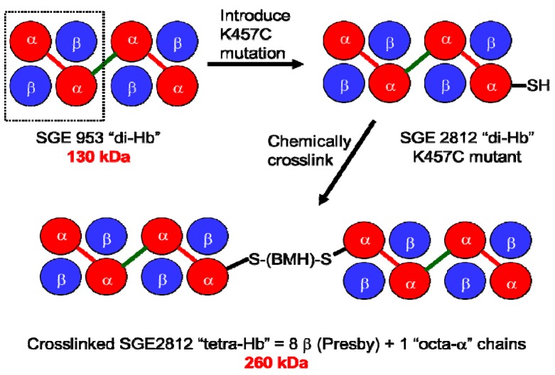
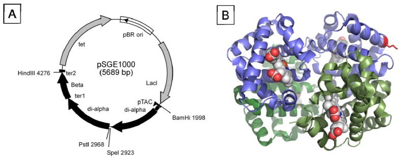
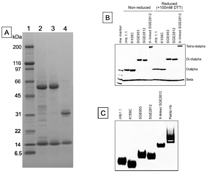
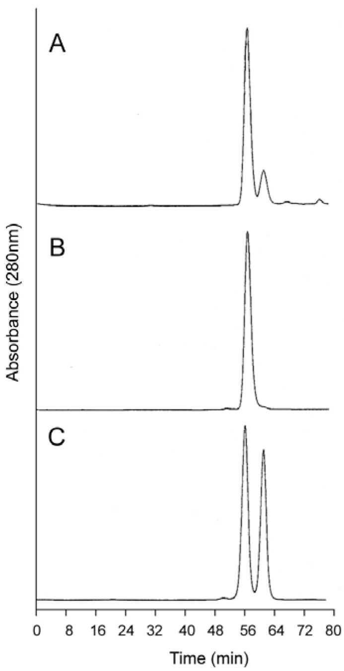
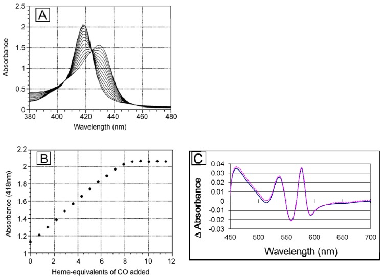
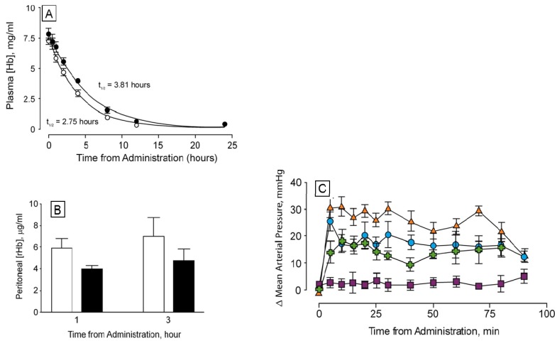
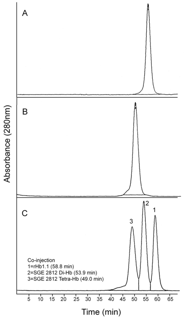
A recombinant 130 kDa dihemoglobin which is made up of a single-chain tetra-α globin and four β globins has been expressed as a soluble protein in E. coli. The sequence of the single chain tetra-α is: αI-Gly-αII-(SerGlyGly)5Ser-αIII-Gly-αIV. This dihemoglobin has been purified and characterized in vitro by size exclusion chromatography, electrospray mass spectroscopy, equilibrium oxygen binding, and analytical ultracentrifugation. The observed values of P50 and nmax for the dihemoglobin are slightly lower than those observed for the recombinant hemoglobin rHb1.1 (a "monohemoglobin" comprised of two β globins and an αI-Gly-αII diα-globin chain). Titration of the deoxy form of dihemoglobin with CO shows that all eight heme centers bind ligand. In vivo, dihemoglobin showed increased circulating halflife and a reduced pressor response in conscious rats when compared to rHb1.1. These observations suggest that dihemoglobin is an oxygen carrying molecule with desirable in vivo properties and provides a platform for an isooncotic hemoglobin solution derived solely from a recombinant source. A 260 kDa tetrahemoglobin has also been produced by chemical crosslinking of a dihemoglobin that contains a Lys16Cys mutation in the C-terminal α-globin subunit. Tetrahemoglobin also shows reduced vasoactivity in conscious rats that is comparable to that observed for dihemoglobin.
Figures
Similar articles
-
Turnover of recombinant human hemoglobin in Escherichia coli occurs rapidly for insoluble and slowly for soluble globin.Arch Biochem Biophys. 1997 Dec 15;348(2):337-46. doi: 10.1006/abbi.1997.0410. Arch Biochem Biophys. 1997. PMID: 9434746
-
Assessing the relative stabilities of engineered hemoglobins using electrospray mass spectrometry.Anal Biochem. 1999 Jul 15;272(1):8-18. doi: 10.1006/abio.1999.4140. Anal Biochem. 1999. PMID: 10405287
-
Expression of fully functional tetrameric human hemoglobin in Escherichia coli.Proc Natl Acad Sci U S A. 1990 Nov;87(21):8521-5. doi: 10.1073/pnas.87.21.8521. Proc Natl Acad Sci U S A. 1990. PMID: 2236062 Free PMC article.
-
Stabilization of apoglobin by low temperature increases yield of soluble recombinant hemoglobin in Escherichia coli.Appl Environ Microbiol. 1997 Nov;63(11):4313-20. doi: 10.1128/aem.63.11.4313-4320.1997. Appl Environ Microbiol. 1997. PMID: 9361418 Free PMC article.
-
Enhancing stability and expression of recombinant human hemoglobin in E. coli: Progress in the development of a recombinant HBOC source.Biochim Biophys Acta. 2008 Oct;1784(10):1471-9. doi: 10.1016/j.bbapap.2008.04.012. Epub 2008 May 1. Biochim Biophys Acta. 2008. PMID: 18489914 Review.
Cited by
-
Comparison of the Pharmacokinetic Properties of Hemoglobin-Based Oxygen Carriers.J Funct Biomater. 2017 Mar 18;8(1):11. doi: 10.3390/jfb8010011. J Funct Biomater. 2017. PMID: 28335469 Free PMC article. Review.
References
-
- Winslow R.M. Blood Substitutes. Academic Press; London, UK: 2006.
-
- Estep T., Bucci E., Farmer M., Greenburg G., Harrington J., Kim H.W., Klein H., Mitchell P., Nemo G., Olsen K., Palmer A., Valeri C.R., Winslow R. Basic science focus on blood substitutes: A summary of the NHLBI Division of Blood Diseases and Resources Working Group Workshop, March 1, 2006. Transfusion. 2008;48:776–782. doi: 10.1111/j.1537-2995.2007.01604.x. - DOI - PubMed
LinkOut - more resources
Full Text Sources
Research Materials
