. 2012 Mar 22;3(1):209-24.
doi: 10.3390/jfb3010209.

Finite-element modeling of viscoelastic cells during high-frequency cyclic strain

Affiliations

Finite-element modeling of viscoelastic cells during high-frequency cyclic strain

Jaques S Milner et al. J Funct Biomater. .

Abstract

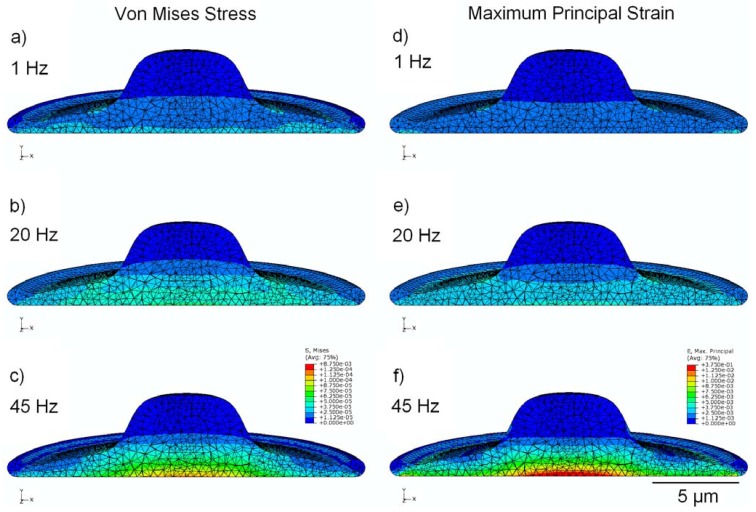
Mechanotransduction refers to the mechanisms by which cells sense and respond to local loads and forces. The process of mechanotransduction plays an important role both in maintaining tissue viability and in remodeling to repair damage; moreover, it may be involved in the initiation and progression of diseases such as osteoarthritis and osteoporosis. An understanding of the mechanisms by which cells respond to surrounding tissue matrices or artificial biomaterials is crucial in regenerative medicine and in influencing cellular differentiation. Recent studies have shown that some cells may be most sensitive to low-amplitude, high-frequency (i.e., 1-100 Hz) mechanical stimulation. Advances in finite-element modeling have made it possible to simulate high-frequency mechanical loading of cells. We have developed a viscoelastic finite-element model of an osteoblastic cell (including cytoskeletal actin stress fibers), attached to an elastomeric membrane undergoing cyclic isotropic radial strain with a peak value of 1,000 µstrain. The results indicate that cells experience significant stress and strain amplification when undergoing high-frequency strain, with peak values of cytoplasmic strain five times higher at 45 Hz than at 1 Hz, and peak Von Mises stress in the nucleus increased by a factor of two. Focal stress and strain amplification in cells undergoing high-frequency mechanical stimulation may play an important role in mechanotransduction.


Figures

Figure 1
Figure 1
(a) Schematic representation of the 3D model geometry used to represent an osteoblastic cell (grey), attached to an elastomeric substrate (gold); (b) Cross-sectional view of the 3D mesh, showing the location and shape of the nucleus (green) within the cytoplasm.
Figure 2
Figure 2
Schematic representation of the 3D model geometry used to represent a network of stress fibers, shown in: (a) top; (b) oblique; and (c) side view.
Figure 3
Figure 3
finite-element modeling (FEM) simulation results, showing peak values of stress in MPa (a,b,c) and strain (d,e,f) over the stimulation cycle, for frequencies of 1 Hz, 20 Hz, and 45 Hz. Cytoplasm is assumed to be viscoelastic, with the elastic modulus of the nucleus four times stiffer.
Figure 4
Figure 4
The impact of the assumed viscoelastic relaxation time constant τ at various frequencies. (a) shows the percentage increase in maximum cytoplasmic stress as a function of frequency, for τ = 0.1, 1 and 10 s, as well as for a linear elastic material; (b) shows the decrease in observed maximum cytoplasmic stress at 1, 20 and 45 Hz as a function of viscoelastic time constant, where the difference is with respect to a linear elastic model.
Figure 5
Figure 5
FEM simulation results under the assumption that both cytoplasm and nucleus are linear isoelastic materials, with elastic modulus of 6.5 kPa and Poisson’s ratio 0.5, showing peak values of stress in MPa (a,b,c) and strain (d,e,f) over the stimulation cycle, for frequencies of 1 Hz, 20 Hz, and 45 Hz.
Figure 6
Figure 6
FEM simulation results, showing peak values of stress in MPa (a,b,c) and strain (d,e,f) over the stimulation cycle, for frequencies of 1 Hz, 20 Hz, and 45 Hz. This model incorporates cytoskeletal elements, represented by 24 pre-tensioned stress fibers.

Similar articles

Cited by

References

    1. Hahn C., Schwartz M.A. Mechanotransduction in vascular physiology and atherogenesis. Nat. Rev. Mol. Cell. Biol. 2009;10:53–62. doi: 10.1038/nrm2596. - DOI - PMC - PubMed
    1. Jaalouk D.E., Lammerding J. Mechanotransduction gone awry. Nat. Rev. Mol. Cell. Biol. 2009;10:63–73. doi: 10.1038/nrm2597. - DOI - PMC - PubMed
    1. Huang C., Ogawa R. Mechanotransduction in bone repair and regeneration. FASEB J. 2010;24:3625–3632. doi: 10.1096/fj.10-157370. - DOI - PubMed
    1. Penley N.J., Schafer C.P., Bartoe J.D. The international space station as a microgravity research platform. Acta Astronaut. 2002;50:691–696. doi: 10.1016/S0094-5765(02)00003-6. - DOI - PubMed
    1. Rogers M.J., Hrovat K., Moskowitz M.E. Effects of exercise equipment on the microgravity environment. Adv. Space. Res. 1999;24:1283–1287. - PubMed

LinkOut - more resources