. 2014 Jun 24:5:4178.
doi: 10.1038/ncomms5178.

IP6K structure and the molecular determinants of catalytic specificity in an inositol phosphate kinase family

Affiliations

IP6K structure and the molecular determinants of catalytic specificity in an inositol phosphate kinase family

Huanchen Wang et al. Nat Commun. .

Abstract

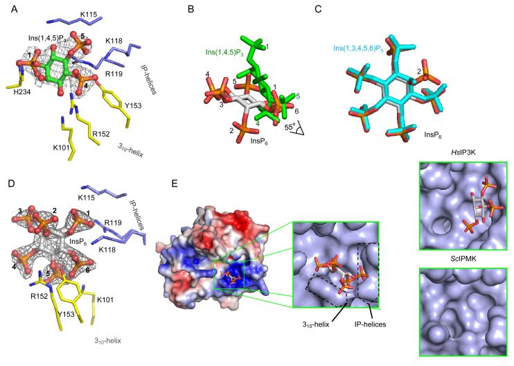
Inositol trisphosphate kinases (IP3Ks) and inositol hexakisphosphate kinases (IP6Ks) each regulate specialized signalling activities by phosphorylating either InsP3 or InsP6 respectively. The molecular basis for these different kinase activities can be illuminated by a structural description of IP6K. Here we describe the crystal structure of an Entamoeba histolytica hybrid IP6K/IP3K, an enzymatic parallel to a 'living fossil'. Through molecular modelling and mutagenesis, we extrapolated our findings to human IP6K2, which retains vestigial IP3K activity. Two structural elements, an α-helical pair and a rare, two-turn 310 helix, together forge a substrate-binding pocket with an open clamshell geometry. InsP6 forms substantial contacts with both structural elements. Relative to InsP6, enzyme-bound InsP3 rotates 55° closer to the α-helices, which provide most of the protein's interactions with InsP3. These data reveal the molecular determinants of IP6K activity, and suggest an unusual evolutionary trajectory for a primordial kinase that could have favored efficient bifunctionality, before propagation of separate IP3Ks and IP6Ks.


Figures

Fig. 1
Fig. 1. HPLC and NMR analysis of the products of Ins(1,4,5)P3 and InsP6 phosphorylation by EhIP6KA
A, HPLC analysis (Partisphere SAX, Gradient 1; see Experimental Procedures) of InsP6 phosphorylation by either EhIP6KA (57 ng, 20 min; solid line) or HsIP6K2 (7.6 ng, 20 min; dotted line). B, NMR analysis of the products of the reaction between InsP6 and ATP catalyzed by EhIP6KA; the Fig. depicts an overlay of the 1D 1H and 2D 1H/31P HSQC spectra. The acquisition parameters are given under Experimental Procedures. Panel C, HPLC analysis (Partisphere SAX, Gradient 2; see Experimental Procedures) of the phosphorylation of approx. 9000 D.P.M. [14C]Ins(1,3,4,5,6)P5 by EhIP6KA (6 μg, 20 min, 50 μl; filled circles). Also shown are the elution of [3H]-labeled internal standards of PP-InsP4 and [PP]2-InsP3, as well as InsP6 (open circles). Panels D and E depict representative HPLC assays (90 min; 200 μl) that contained approx. 10,000 D.P.M. [3H]Ins(1,4,5)P3 plus 2 μg EhIP6KA. After quenching and neutralization, products (filled circles) were analyzed by Q100 SAX HPLC (Gradient 3). Panel D includes the elution positions of standards of [3H]Ins(1,2,4,5)P4 and [3H]InsP6 (open circles), determined in parallel HPLC runs. Panel E zooms in on the InsP4 region of the chromatograph; elution of internal [14C] standards of Ins(1,3,4,5)P4 and Ins(1,4,5,6)P4 are also shown (open circles).
Fig. 2
Fig. 2. Overall structure of EhIP6KA
A, Ribbon-plot of EhIP6KA structure. ATP and InsP6 are shown as sticks within a transparent surface. Two Mg atoms are depicted as magenta balls. B, Topology diagram. C, A manual alignment of amino-acid sequences (EhIP6KA, XP-648490.2; HsIP6K2, NP_057375.2; ScIPMK, NP_010458.3; HsIP3KA, NP_002211.1), guided by the structural elements that have been observed in crystal structures, and in the case of HsIP6K2, secondary structural predictions. Outside of the conserved catalytic core of the HsIP6K2 are two significant insertions that are omitted from the alignment. The first of these (residues 73-202) includes a specialized HSP90-binding domain . The second insertion (residues 340-371) includes a CK2-regulated, ubiquitination motif . The secondary structural elements from EhIP6KA are depicted above its sequence and are color-coded orange for the N-lobe, yellow for the C-lobe and blue for the IP-helix. Structural elements that directly participate in substrate interactions are highlighted by shading. Residues in HsIP6K2 that were selected for mutagenesis are colored red. PDB codes for EhIP6KA are 4O4B, 4O4C, 4O4D, 4O4E, 4O4F.
Fig. 3
Fig. 3. Nucleotide binding by EhIP6KA
A, ATP is depicted as a stick and ball model. Two Mg atoms are depicted as magenta spheres. Polar contacts are shown with dashed lines. Amino acids are shown as stick. B, Metal coordination. Two Mg atoms are depicted as magenta spheres. Water molecules are depicted as red spheres. The structure of ATP and Asp231are shown as stick models. Polar contacts to coordinate with Mg atoms are shown with dashed lines. C, The orientation of the EhIP6KA-bound nucleotide (green for carbon, red for oxygen, blue for nitrogen and orange for phosphorus atoms) and Mg atoms (magenta spheres) are superimposed upon that for HsIP3KA (grey stick represent AMPPNP (ATP analog); grey spheres represent Mg), and ScIPMK (light blue stick and spheres represent ADP and Mg).
Fig. 4
Fig. 4. Ligplots showing interactions of EhIP6KA with ATP and inositol phosphates
Ligplots are shown for A, ATP and Ins(1,4,5)P3 and B, InsP6. Hydrogen bonds are shown in green dashed lines; bond distances are denoted in Angstroms. Residues that make hydrophobic interactions are depicted as grey eyelashes (cutoff distance is 3.9 Å). Atoms are shown white for carbon, red for oxygen, blue for nitrogen and orange for phosphorus.
Fig. 5
Fig. 5. Inositol phosphate binding site for EhIP6KA
A, Binding of Ins(1,4,5)P3 (stick and ball model; green for carbon, red for oxygen and orange for phosphorus atoms. The phosphate groups are numbered). The refined 2Fo-Fc electron density map is contoured at 1.3 σ. B, Overlay of InsP6 (grey stick model with orange and red phosphate groups) and Ins(1,4,5)P3 (green stick). C, Overlay of InsP6 and Ins(1,3,4,5,6)P5 (cyan stick). D, Binding of InsP6 (stick and ball model; green for carbon, red for oxygen and orange for phosphorus atoms). The phosphate groups are numbered. Amino acids are shown as stick models. The refined 2Fo-Fc electron density map is contoured at 1.3 σ. E, Electrostatic surface plot with blue and red coloration to respectively indicate positive and negative electrostatic potentials at physiological pH. In the magnified surface representation, the positions of the 310 helix and IP-helices are highlighted. Bound InsP6 is depicted as a stick model. Also shown are surface representation of Ins(1,4,5)P3 in the active site of HsIP3K (data from ) and the active site of ScIPMK (data from ; crystals did not contain substrate).
Fig. 6
Fig. 6. HPLC analysis of the kinase activities of wild-type and mutant IP6Ks
A, InsP6 kinase (black bars) and Ins(1,4,5)P3 kinase (grey bars) activities of wild-type HsIP6K2 and the indicated mutants (means ± standard errors, n ≥3). Panels B shows representative HPLC analysis (Partisphere SAX; Gradient 2) of Ins(1,4,5)P3 kinase activities of wild-type HsIP6K2 (8 μg, 7 hr). The inset to panel B shows HPLC analysis (Q100 SAX; Gradient 3) of the InsP4 products formed by the wild-type enzyme (closed circles) together with the elution of internal standards of [14C]-Ins(1,3,4,5)P4 and [14C]-Ins(1,4,5,6)P4 (open circles). Panel C shows a representative HPLC analysis (Partisphere SAX; Gradient 2) of Ins(1,4,5)P3 kinase activity of a Cys257Glu mutant of HsIP6K2 (5 μg, 1 hr). Panel D shows a representative HPLC analysis (Q100 SAX; Gradient 3) of the InsP4 products formed by a Cys257Glu mutant of HsIP6K2 (5 μg, 45 min). Panel E shows a representative HPLC analysis of Ins(1,4,5)P3 kinase activity of a Gln240Met mutant of HsIP6K2 (11 μg, 7 hr).

Similar articles

Cited by

References

    1. Shears SB. Diphosphoinositol polyphosphates: metabolic messengers? Mol. Pharmacol. 2009;76:236–252. - PMC - PubMed
    1. Wilson MS, Livermore TM, Saiardi A. Inositol pyrophosphates: between signalling and metabolism. Biochem. J. 2013;452:369–379. - PubMed
    1. Szijgyarto Z, Garedew A, Azevedo C, Saiardi A. Influence of inositol pyrophosphates on cellular energy dynamics. Science. 2011;334:802–805. - PubMed
    1. Illies C, et al. Inositol pyrophosphates determine exocytic capacity. Science. 2007;318:1299–1302. - PubMed
    1. Chakraborty A, et al. Inositol pyrophosphates inhibit AKT signaling, thereby regulating insulin sensitivity and weight gain. Cell. 2010;143:897–910. - PMC - PubMed

Publication types

MeSH terms

Substances

LinkOut - more resources