

Detection of sickle cell hemoglobin in Haiti by genotyping and hemoglobin solubility tests
- PMID: 24957539
- PMCID: PMC4125270
- DOI: 10.4269/ajtmh.13-0572
Detection of sickle cell hemoglobin in Haiti by genotyping and hemoglobin solubility tests
Abstract
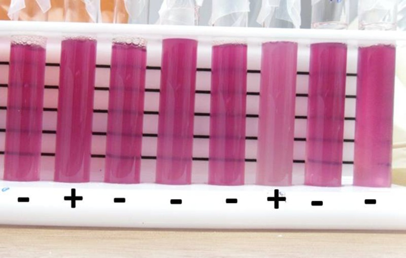
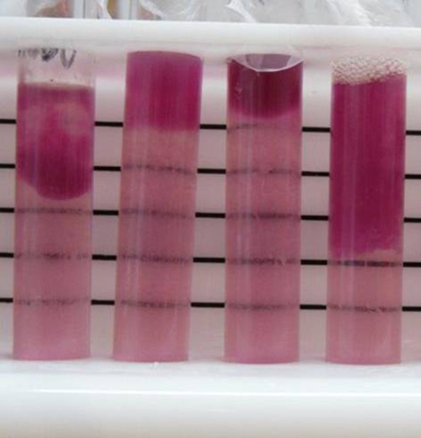
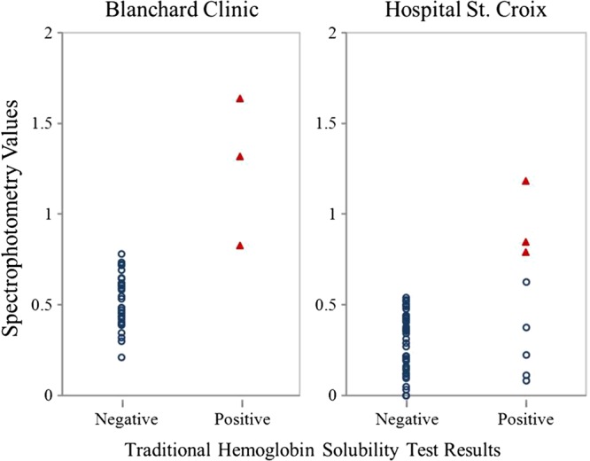
Sickle cell disease is a growing global health concern because infants born with the disorder in developing countries are now surviving longer with little access to diagnostic and management options. In Haiti, the current state of sickle cell disease/trait in the population is unclear. To inform future screening efforts in Haiti, we assayed sickle hemoglobin mutations using traditional hemoglobin solubility tests (HST) and add-on techniques, which incorporated spectrophotometry and insoluble hemoglobin separation. We also generated genotype data as a metric for HST performance. We found 19 of 202 individuals screened with HST were positive for sickle hemoglobin, five of whom did not carry the HbS allele. We show that spectrophotometry and insoluble hemoglobin separation add-on techniques could resolve false positives associated with the traditional HST approach, with some limitations. We also discuss the incorporation of insoluble hemoglobin separation observation with HST in suboptimal screening settings like Haiti.
© The American Society of Tropical Medicine and Hygiene.
Figures
Similar articles
-
Novel test method (sickle confirm) to differentiate sickle cell anemia from sickle cell trait for potential use in developing countries.Clin Lab Sci. 2012 Winter;25(1):26-34. Clin Lab Sci. 2012. PMID: 22458046
-
Trimodal distribution of HbS levels in sickle heterozygotes--an useful predictor of the alpha-genotype for population screening.Indian J Med Res. 1998 Dec;108:285-90. Indian J Med Res. 1998. PMID: 9919012 Clinical Trial.
-
Sickle hemoglobin (HbS) allele and sickle cell disease: a HuGE review.Am J Epidemiol. 2000 May 1;151(9):839-45. doi: 10.1093/oxfordjournals.aje.a010288. Am J Epidemiol. 2000. PMID: 10791557 Review.
-
Non-enzymatic digoxigenin labeling of allele-specific oligonucleotides for the detection of the sickle cell mutation.Eur J Haematol. 1992 Nov;49(5):275-6. doi: 10.1111/j.1600-0609.1992.tb00061.x. Eur J Haematol. 1992. PMID: 1473589 No abstract available.
-
Review: the sickle hemoglobinopathies--genetic analyses of common phenocopies and new molecular approaches to treatment.Am J Med Sci. 1984 Nov;288(4):169-74. doi: 10.1097/00000441-198411000-00004. Am J Med Sci. 1984. PMID: 6208780 Review.
Cited by
-
Determinants of Anemia and Hemoglobin Concentration in Haitian School-Aged Children.Am J Trop Med Hyg. 2015 Nov;93(5):1092-8. doi: 10.4269/ajtmh.15-0073. Epub 2015 Sep 8. Am J Trop Med Hyg. 2015. PMID: 26350448 Free PMC article. Clinical Trial.
-
Genetic Diversity of Plasmodium falciparum in Haiti: Insights from Microsatellite Markers.PLoS One. 2015 Oct 13;10(10):e0140416. doi: 10.1371/journal.pone.0140416. eCollection 2015. PLoS One. 2015. PMID: 26462203 Free PMC article.
-
Investigation of mutations in the HBB gene using the 1,000 genomes database.PLoS One. 2017 Apr 5;12(4):e0174637. doi: 10.1371/journal.pone.0174637. eCollection 2017. PLoS One. 2017. PMID: 28379995 Free PMC article.
References
-
- Ingram VM. Gene mutations in human hemoglobin: the chemical difference between normal and sickle cell hemoglobin. Nature. 1957;180:326–328. - PubMed
-
- Ashley-Koch A, Yang Q, Olney RS. Sickle hemoglobin (HbS) allele and sickle cell disease: a HuGE review. Am J Epidemiol. 2000;151:839–845. - PubMed
-
- Hunt JA, Ingram VM. Abnormal human hemoglobins. IV. The chemical difference between normal human hemoglobin and hemoglobin C. Biochim Biophys Acta. 1960;42:409–421. - PubMed
-
- Rees DC, Williams TN, Gladwin MT. Sickle-cell disease. Lancet. 2010;376:2018–2031. - PubMed
Publication types
MeSH terms
Substances
LinkOut - more resources
Full Text Sources
Other Literature Sources
Medical
