. 2014 Sep;91(3):589-97.
doi: 10.4269/ajtmh.14-0146. Epub 2014 Jun 23.

Phylogeography of Rickettsia rickettsii genotypes associated with fatal Rocky Mountain spotted fever

Affiliations

Phylogeography of Rickettsia rickettsii genotypes associated with fatal Rocky Mountain spotted fever

Christopher D Paddock et al. Am J Trop Med Hyg. 2014 Sep.

Abstract

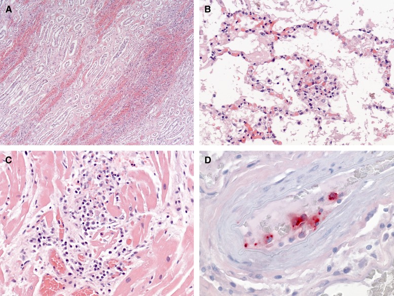
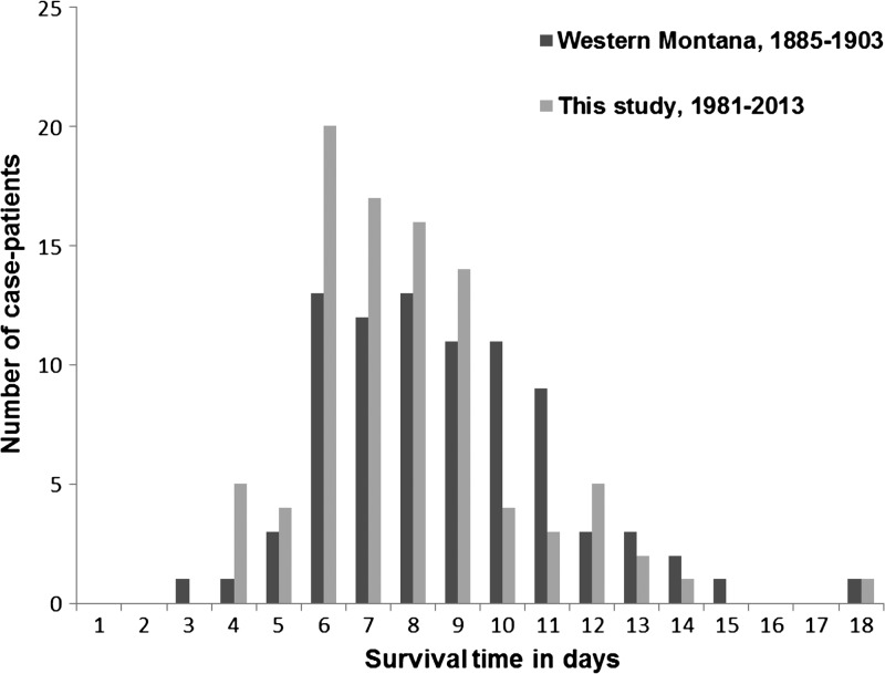
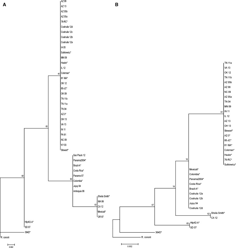
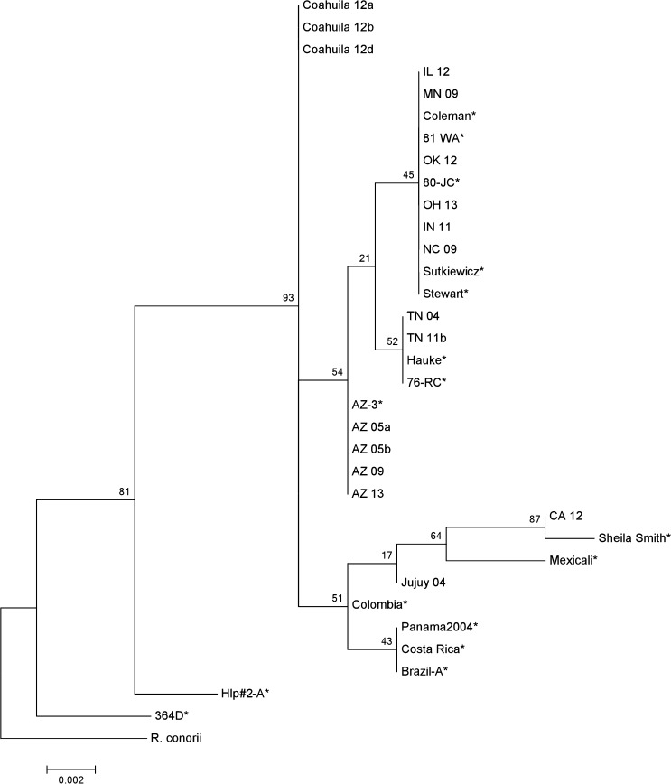
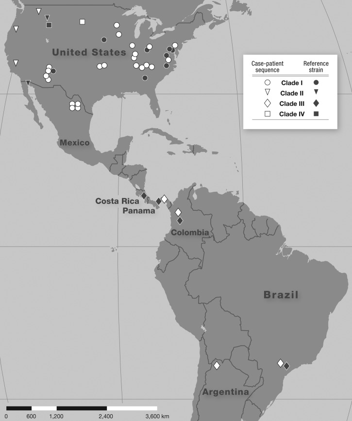
Rocky Mountain spotted fever (RMSF), a tick-borne zoonosis caused by Rickettsia rickettsii, is among the deadliest of all infectious diseases. To identify the distribution of various genotypes of R. rickettsii associated with fatal RMSF, we applied molecular typing methods to samples of DNA extracted from formalin-fixed, paraffin-embedded tissue specimens obtained at autopsy from 103 case-patients from seven countries who died of RMSF. Complete sequences of one or more intergenic regions were amplified from tissues of 30 (29%) case-patients and revealed a distribution of genotypes consisting of four distinct clades, including the Hlp clade, regarded previously as a non-pathogenic strain of R. rickettsii. Distinct phylogeographic patterns were identified when composite case-patient and reference strain data were mapped to the state and country of origin. The phylogeography of R. rickettsii is likely determined by ecological and environmental factors that exist independently of the distribution of a particular tick vector.


Figures

Figure 1.
Figure 1.
Survival times (dark bars, median = 8 days), recorded for 84 fatal cases of Rocky Mountain spotted fever (RMSF) from western Montana during 1885–19031 compared with survival times of 92 case-patients with fatal RMSF from North, Central, and South America, evaluated during 1981–2013 (light bars, median = 8 days). One case-patient from the Montana series who died on the 27th day was excluded from the comparison.
Figure 2.
Figure 2.
Histopathology and immununohistochemical staining of various tissues from a patient with fatal Rocky Mountain spotted fever caused by an Hlp genotype of Rickettsia rickettsii, including interstitial nephritis (A), pulmonary capillaritis and intra-alveolar edema (B), and lymphohistiocytic myocarditis (C). Immunohistochemical staining of intracellular R. rickettsii antigens (red) in endothelial cells in a vessel in the brainstem (D). Hematoxylin and eosin stain (AC), and immunoalkaline phosphatase staining technique using a hyperimmune rabbit anti-R. rickettsii antiserum with naphthol-fast red and hematoxylin counterstain (D). Original magnifications ×12.5 (A), ×50 (B and C), and ×100 (D).
Figure 3.
Figure 3.
Dendrogram showing phylogenetic relationships of Rickettsia rickettsii RR0155-rpmB (A) and RR1240-tlc5 (B) sequences, obtained from formalin-fixed and paraffin-embedded tissues of patients with fatal Rocky Mountain spotted fever (RMSF). Previously evaluated reference strains of R. rickettsii, isolated predominantly from patients with fatal RMSF, are marked by asterisks. Bootstrap values (1,000 replicates) are shown above the nodes. Rickettsia conorii represents the out-group. The scale bar corresponds to the number of steps.
Figure 4.
Figure 4.
Dendrogram comprising concatenated sequences of RR0155-rpmB, cspA-ksgA, and RR1240-tlc5 sequences from formalin-fixed and paraffin-embedded tissue sections of 17 patients with fatal Rocky Mountain spotted fever. Previously evaluated reference strains of Rickettsia rickettsii, isolated predominantly from patients with fatal RMSF, are marked by asterisks. Bootstrap values (1,000 replicates) are shown above the nodes. Rickettsia conorii represents the out-group. The scale bar corresponds to the number of steps.
Figure 5.
Figure 5.
Geographic distribution of genetic clades of Rickettsia rickettsii (white symbols) identified in formalin-fixed and paraffin-embedded tissues of 30 patients with fatal Rocky Mountain spotted fever from Argentina, Brazil, Colombia, Mexico, Panama, and the United States, 1991–2013. Black symbols represent the locations of origin for previously characterized reference strains from which the clade designations were determined., Domestic locations are mapped to the county level, whereas international locations have been mapped to the state, province, or department level.

References

    1. Wilson LB, Chowning WM. Studies in Piroplasma hominis. (Spotted fever or “tick fever” of the Rocky Mountains) J Infect Dis. 1904;1:31–57. - PubMed
    1. Philip RN. Rocky Mountain Spotted Fever in Western Montana: Anatomy of a Pestilence. Hamilton, MT: Bitter Root Valley Historical Society; 2000. pp. 55–69.
    1. Maxey EE. Some observations on the so-called spotted fever of Idaho. Med Sentinel. 1899;7:432–438.
    1. Spencer WO. Mountain or spotted fever, as seen in Idaho and eastern Oregon. Med Sentinel. 1907;15:532–537.
    1. Ricketts HT. Some aspects of Rocky Mountain spotted fever as shown by recent investigations. Med Record. 1909;76:843–855. - PubMed

Publication types

MeSH terms

LinkOut - more resources