. 2014 Jul;42(13):8330-42.
doi: 10.1093/nar/gku551. Epub 2014 Jun 23.

CstF-64 supports pluripotency and regulates cell cycle progression in embryonic stem cells through histone 3' end processing

Affiliations

CstF-64 supports pluripotency and regulates cell cycle progression in embryonic stem cells through histone 3' end processing

Bradford A Youngblood et al. Nucleic Acids Res. 2014 Jul.

Abstract

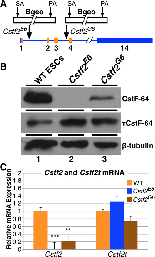
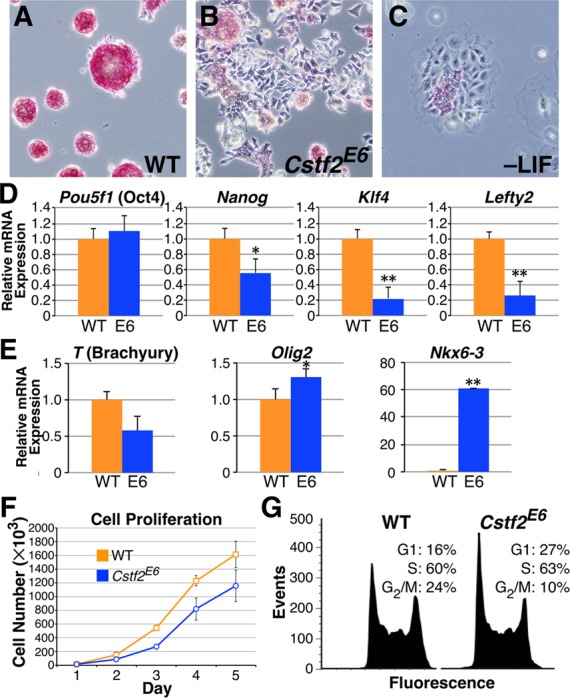
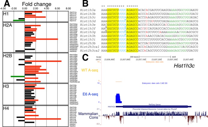
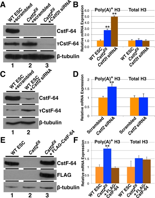
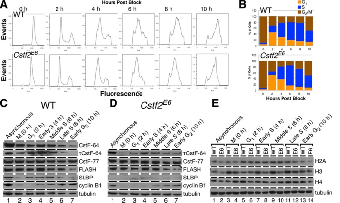
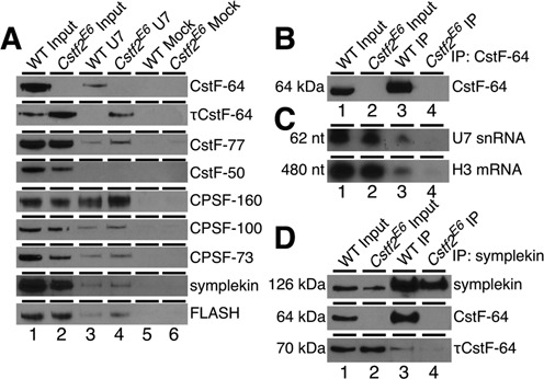
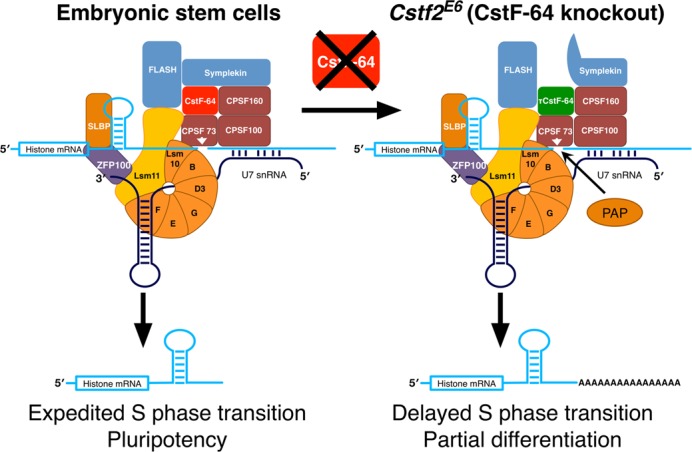
Embryonic stem cells (ESCs) exhibit a unique cell cycle with a shortened G1 phase that supports their pluripotency, while apparently buffering them against pro-differentiation stimuli. In ESCs, expression of replication-dependent histones is a main component of this abbreviated G1 phase, although the details of this mechanism are not well understood. Similarly, the role of 3' end processing in regulation of ESC pluripotency and cell cycle is poorly understood. To better understand these processes, we examined mouse ESCs that lack the 3' end-processing factor CstF-64. These ESCs display slower growth, loss of pluripotency and a lengthened G1 phase, correlating with increased polyadenylation of histone mRNAs. Interestingly, these ESCs also express the τCstF-64 paralog of CstF-64. However, τCstF-64 only partially compensates for lost CstF-64 function, despite being recruited to the histone mRNA 3' end-processing complex. Reduction of τCstF-64 in CstF-64-deficient ESCs results in even greater levels of histone mRNA polyadenylation, suggesting that both CstF-64 and τCstF-64 function to inhibit polyadenylation of histone mRNAs. These results suggest that CstF-64 plays a key role in modulating the cell cycle in ESCs while simultaneously controlling histone mRNA 3' end processing.


Figures

Figure 1.
Figure 1.
Expression of CstF-64 and τCstF-64 in wild type and Cstf2 gene-trap interrupted Cstf2E6 and Cstf2G6 mouse embryonic stem cells. (A) Schematic representation of insertion of the gene-trap β-galactosidase-neomycin (Bgeo) fusion protein in the first (Cstf2E6) and third (Cstf2G6) introns of the Cstf2 gene in the two respective ESC lines. The gene-trap consists of a splice acceptor (SA) site and polyadenylation (PA) signal. The lighter shade represents the open reading frame of Cstf2 mRNA. (B) Western blot analysis of CstF-64 and τCstF-64 expression in WT (lane 1), Cstf2E6 (lane 2) and Cstf2G6 ESCs (lane 3). (C) Relative mRNA expression analysis of Cstf2 and Cstf2t mRNAs in the wild type, Cstf2E6 and Cstf2G6 ESC lines. ** denotes P < 0.01 and *** denotes P < 0.001.
Figure 2.
Figure 2.
Loss of CstF-64 results in diminished pluripotent state, decreased cell proliferation and disrupted cell cycle. Alkaline phosphatase staining of (A) WT ESCs, (B) Cstf2E6 ESCs and (C) WT ESCs cultured without LIF for 96 h, 100X magnification. (D) Relative mRNA expression of the pluripotency regulators, Pou5f1 (Oct4), Nanog, Klf4 and Lefty2 and (E) differentiation markers, T (Brachyury), Olig2 and Nkx6-3, representing mesoderm, ectoderm and endoderm germ layers, respectively. * denotes P < 0.05 and ** denotes P < 0.01. (F) Growth rate analysis of WT and Cstf2E6 ESCs. Cells were plated in triplicate and data points represent the average cell count. Standard deviation is also shown. The growth rates on days 3, 4 and 5 between the WT and Cstf2E6 ESCs were statistically significant at P < 0.05 using a one-tailed t-test. (G) Cell cycle analysis of WT and Cstf2E6 ESCs stained with PI and analyzed using flow cytometry. For the cell cycle analysis, a two-tailed t-test was performed to obtain significance on triplicate samples. The P values for both G1 and G2/M phases is P < 0.05.
Figure 3.
Figure 3.
Increase in the polyadenylation of replication-dependent histone mRNAs in Cstf2E6 cells. (A) Fold change of the polyadenylated replication-dependent histones families in the Cstf2E6 ESCs versus wild type ESCs. Green bars indicate 2-fold or more down-regulation, red bars—2-fold or more up-regulation and black bars no change (<2-fold) of the expression of the polyadenylated histone mRNAs. (B) Cleavage and polyadenylation sites in the replication-dependent histone H3 family. Alignment of the stem-loop region, 3′ end processing cleavage site and downstream genomic sequences. Red nucleotides indicate the 3′ end cleavage and polyadenylation sites as determined by A-seq in wild type ESCs and Cstf2E6 cells. The stem-loop region of the H3 histone mRNAs are highlighted in yellow and blue, respectively. Underlined nucleotides indicate the different nucleotides from the consensus sequence. Asterisks at the top of the alignment point out to the consensus sequence of the stem-loop. Green nucleotides show the respective HDE sequence (AAAGAGCUGU). (C) A-seq reads mapped to the mouse genome (mm9) for wild type and Cstf2E6 ESCs. Position of the Hist1h3c is shown in the RefSeq Genes track. The blue peak above the RefSeq Genes track is the number of non-normalized reads uniquely mapping to histone Hist1h3c obtained from A-seq for the Cstf2E6 cells overlapping with the stem-loop region. Above in orange—A-seq reads obtained from the wild type ESCs. The sequence conservation of Hist1h3c in placental mammals is shown at the bottom of the figure.
Figure 4.
Figure 4.
CstF-64 and τCstF-64 are necessary for normal 3′ end processing of replication-dependent histone mRNAs. (A) Western blot analysis of CstF-64 and τCstF-64 expression in WT (lane 1), Cstf2E6 ESCs transfected with scrambled siRNA (lane 2) and Cstf2E6 ESCs transfected with siRNA specific for Cstf2t gene (lane 3). (B) Relative mRNA expression analysis of polyadenylated and total Hist1h3c histone mRNA in wild type ESCs (orange), Cstf2E6 cells transfected with scrambled siRNA (blue) or Cstf2E6 cells transfected with siRNA against Cstf2t gene (brown). (C) Western blot analysis of CstF-64 and τCstF-64 expression in WT ESCs either transfected with scrambled siRNA (lane 1) or Cstf2 gene specific siRNA (lane 2). (D) Relative mRNA expression analysis of polyadenylated and total Hist1h3c histone mRNA in wild type ESCs transfected with scrambled (orange) or Cstf2 gene specific siRNA (blue). (E) Western blot analysis of CstF-64 expression in WT ESCs (lane 1), Cstf2E6 ESCs (lane 2) and Cstf2E6 ESCs ectopically expressing CstF-64 (lane 3). (F) Corresponding relative mRNA expression analysis of polyadenylated and total Hist1h3c histone mRNA. * denotes P < 0.05 and ** P < 0.01.
Figure 5.
Figure 5.
CstF-64 is required for S-phase entry and histone expression. (A) Cell cycle analysis of WT and Cstf2E6 ESCs mitotically synchronized (0 h) with nocodazole and subsequently released for 10 h in 2 h time points (2–10 h). Cells were stained with PI and analyzed using flow cytometry. (B) Corresponding percentages of synchronized and released WT and Cstf2E6 ESCs in the cell cycle phases, G1 (orange), S (blue) and G2/M (brown). (C, D) Western blot analysis detecting the expression of CstF-64, τCstF-64, CstF-77 and the histone processing components, FLASH and SLBP in mitotically synchronized WT and Cstf2E6 ESCs that were released for 10 h post block. Cyclin B1 expression indicates G2/M phase transition. (E) Comparative western blot analysis of the core histone families, H2B, H3 and H4 in mitotically synchronized WT and Cstf2E6 ESCs.
Figure 6.
Figure 6.
CstF-64 is a component of the replication-dependent histone mRNA 3′ end-processing complex. (A) Western blot analysis of the proteins isolated from a pull-down experiment using anti-U7 snRNP oligonucleotide (lanes 3 and 4) or unrelated mock oligonucleotide (lanes 5 and 6) in WT and Cstf2E6 ESCs nuclear extracts. 1/100th of the nuclear extracts from the wild type ESCs (lane 1) and Cstf2E6 cells (lane 2) before the pull down were also loaded on the gel serving as an input control. Antibodies against the indicated proteins were used. (B) Western blot analysis of immunoprecipitation using an anti-CstF-64 antibody in wild type ECCs (lane 3) and Cstf2E6 (lane 4) cells. 1/100th of the total proteins was also loaded as an input control. Wild type ESCs (lane 1) and Cstf2E6 cells (lane 2). (C) Northern blot of RNA from immunoprecipitation with antibodies against CstF-64 that were hybridized with radiolabeled ribo-probes specific for U7 snRNA or Hist1h3c mRNA. Lane 1, 2 μg of total RNA from wild type ESCs; lane 2, 2 μg of total RNA from Cstf2E6 cells; lanes 3–4, 2 μg of RNA purified from CstF-64 immunoprecipitation from wild type ESCs (lane 3) or Cstf2E6 cells (lane 4). (D) Western blot analysis of immunoprecipitation with an anti-symplekin antibody in wild type (lane 3) and Cstf2E6 (lane 4) ESCs protein lysates. IP precipitates from wild type and Cstf2E6 ESCs were probed for interaction with CstF-64 and τCstF-64 as indicated.
Figure 7.
Figure 7.
Schematic representation of how depletion of CstF-64 increases polyadenylation of replication-dependent histone mRNAs and modulates the cell cycle in ESCs and therefore pluripotency. The panel on the left describes the histone mRNA 3′ end processing complex in wild type ESCs. On the right: modified histone mRNA 3′ end processing complex in the Cstf2E6 (CstF-64 knockout) cells. Histone mRNA cleavage occurs in wild type mouse ESCs due to interactions of the U7 snRNP with the histone mRNA downstream element (left panel). Other proteins involved in mRNA polyadenylation further associate with the complex, including CstF-64 and symplekin. The complex promotes site-specific cleavage of the histone mRNA by CPSF-73. Together, these processes correlate with normal entry into S-phase and pluripotency. In normal ESCs, a small amount of these cleaved transcripts are polyadenylated. In Cstf2E6 cells (right panel), CstF-64 is absent, resulting in recruitment of τCstF-64 to the histone 3′ end processing complex (although τCstF-64 interacts more poorly with symplekin). This results in an increase in polyadenylation of the cleaved histone transcripts, presumably by poly(A) polymerase (PAP).

Similar articles

Cited by

References

    1. Pauklin S., Vallier L. The cell-cycle state of stem cells determines cell fate propensity. Cell. 2013;155:135–147. - PMC - PubMed
    1. Coronado D., Godet M., Bourillot P.Y., Tapponnier Y., Bernat A., Petit M., Afanassieff M., Markossian S., Malashicheva A., Iacone R., et al. A short G1 phase is an intrinsic determinant of naive embryonic stem cell pluripotency. Stem Cell Res. 2013;10:118–131. - PubMed
    1. Ghule P.N., Medina R., Lengner C.J., Mandeville M., Qiao M., Dominski Z., Lian J.B., Stein J.L., van Wijnen A.J., Stein G.S. Reprogramming the pluripotent cell cycle: restoration of an abbreviated G1 phase in human induced pluripotent stem (iPS) cells. J. Cell. Physiol. 2011;226:1149–1156. - PMC - PubMed
    1. Becker K.A., Stein J.L., Lian J.B., van Wijnen A.J., Stein G.S. Establishment of histone gene regulation and cell cycle checkpoint control in human embryonic stem cells. J. Cell. Physiol. 2007;210:517–526. - PubMed
    1. Koledova Z., Kafkova L.R., Calabkova L., Krystof V., Dolezel P., Divoky V. Cdk2 inhibition prolongs G1 phase progression in mouse embryonic stem cells. Stem Cells Dev. 2010;19:181–194. - PubMed

Publication types

MeSH terms