. 2012 Feb 27;2(1):91-117.
doi: 10.3390/membranes2010091.

The BAR Domain Superfamily Proteins from Subcellular Structures to Human Diseases

Affiliations

The BAR Domain Superfamily Proteins from Subcellular Structures to Human Diseases

Fatemeh Safari et al. Membranes (Basel). .

Abstract

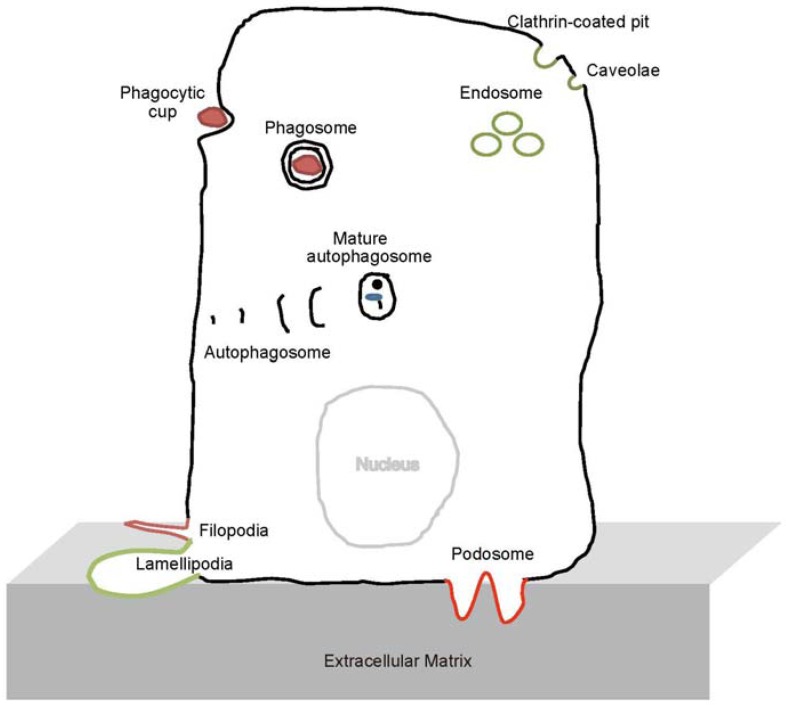
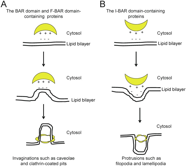
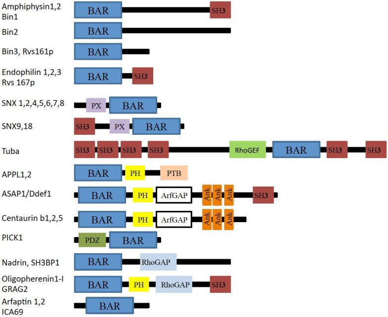
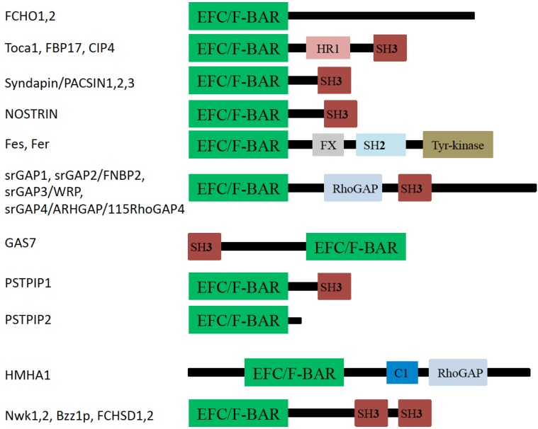
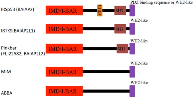
Eukaryotic cells have complicated membrane systems. The outermost plasma membrane contains various substructures, such as invaginations and protrusions, which are involved in endocytosis and cell migration. Moreover, the intracellular membrane compartments, such as autophagosomes and endosomes, are essential for cellular viability. The Bin-Amphiphysin-Rvs167 (BAR) domain superfamily proteins are important players in membrane remodeling through their structurally determined membrane binding surfaces. A variety of BAR domain superfamily proteins exist, and each family member appears to be involved in the formation of certain subcellular structures or intracellular membrane compartments. Most of the BAR domain superfamily proteins contain SH3 domains, which bind to the membrane scission molecule, dynamin, as well as the actin regulatory WASP/WAVE proteins and several signal transduction molecules, providing possible links between the membrane and the cytoskeleton or other machineries. In this review, we summarize the current information about each BAR superfamily protein with an SH3 domain(s). The involvement of BAR domain superfamily proteins in various diseases is also discussed.


Figures

Figure 1
Figure 1
Subcellular structures on which Bin-Amphiphysin-Rvs167 (BAR) domain proteins act, as discussed in this review.
Figure 2
Figure 2
Schematic models for membrane deformation, based on the geometries of basic-charged amino acid residues that correspond to the structures of the membrane-binding surface of the F-BAR domain-containing proteins. (A) BAR or F-BAR proteins bind to the membrane to generate invaginations, such as caveolae and clathrin-coated pits; (B) I-BAR proteins deform the membrane to generate protrusions, such as filopodia and lamellipodia.
Figure 3
Figure 3
Domain organization of BAR subfamily proteins. BAR: Bin-Amphiphysin-Rvs domain; SH3: Src homology 3 domain; RhoGAP: Rho GTPase activating protein domain; PX: Phox homology domain; PH: Pleckstrin Homology domain; ArfGAP: Arf Rho GTPase activating protein domain; PTB: Phospho-tyrosine binding domain; PDZ: Psd-95, Dlg and ZO1 domain; RhoGEF: Rho guanine-nucleotide exchange factors domain; Ank: Ankyrin.
Figure 4
Figure 4
Domain organization of F-BAR proteins. EFC: extended FER-CIP4 homology (EFC) or FCH and BAR (F-BAR) domain; HR1: Protein kinase C-related kinase homology region 1 domain; SH2: Src homology 2 domain; SH3: Src homology 3 domain; FX: F-BAR extension domain; Tyr-kinase: Tyrosine kinase domain; RhoGAP: Rho GTPase activating protein domain; C1: Protein kinase C conserved region 1.
Figure 5
Figure 5
Schematic of domain organization of I-BAR proteins. IRSp53-MIM homology domain (IMD)/inverse-BAR (I-BAR); CRIB: Cdc42-Rac interactive binding region; SH3: Src homology 3 domain; WH2: Wasp homology 2 (verprolin homology) domain.

References

    1. Qualmann B., Koch D., Kessels M.M. Let’s go bananas: Revisiting the endocytic BAR code. EMBO J. 2011;30:3501–3515. doi: 10.1038/emboj.2011.266. - DOI - PMC - PubMed
    1. Suetsugu S., Toyooka K., Senju Y. Subcellular membrane curvature mediated by the BAR domain superfamily proteins. Semin. Cell Dev. Biol. 2010;21:340–349. - PubMed
    1. Frost A., Unger V.M., De Camilli P. The BAR domain superfamily: Membrane-molding macromolecules. Cell. 2009;137:191–196. doi: 10.1016/j.cell.2009.04.010. - DOI - PMC - PubMed
    1. Doherty G.J., McMahon H.T. Mediation, modulation, and consequences of membrane-cytoskeleton interactions. Annu. Rev. Biophys. 2008;37:65–95. doi: 10.1146/annurev.biophys.37.032807.125912. - DOI - PubMed
    1. Takenawa T., Suetsugu S. The WASP-WAVE protein network: Connecting the membrane to the cytoskeleton. Nat. Rev. Mol. Cell Biol. 2007;8:37–48. doi: 10.1038/nrm2069. - DOI - PubMed

LinkOut - more resources