. 2014 Sep;34(17):3259-71.
doi: 10.1128/MCB.00472-14. Epub 2014 Jun 23.

Yeast Dun1 kinase regulates ribonucleotide reductase inhibitor Sml1 in response to iron deficiency

Affiliations

Yeast Dun1 kinase regulates ribonucleotide reductase inhibitor Sml1 in response to iron deficiency

Nerea Sanvisens et al. Mol Cell Biol. 2014 Sep.

Abstract

Iron is an essential micronutrient for all eukaryotic organisms because it participates as a redox-active cofactor in many biological processes, including DNA replication and repair. Eukaryotic ribonucleotide reductases (RNRs) are Fe-dependent enzymes that catalyze deoxyribonucleoside diphosphate (dNDP) synthesis. We show here that the levels of the Sml1 protein, a yeast RNR large-subunit inhibitor, specifically decrease in response to both nutritional and genetic Fe deficiencies in a Dun1-dependent but Mec1/Rad53- and Aft1-independent manner. The decline of Sml1 protein levels upon Fe starvation depends on Dun1 forkhead-associated and kinase domains, the 26S proteasome, and the vacuolar proteolytic pathway. Depletion of core components of the mitochondrial iron-sulfur cluster assembly leads to a Dun1-dependent diminution of Sml1 protein levels. The physiological relevance of Sml1 downregulation by Dun1 under low-Fe conditions is highlighted by the synthetic growth defect observed between dun1Δ and fet3Δ fet4Δ mutants, which is rescued by SML1 deletion. Consistent with an increase in RNR function, Rnr1 protein levels are upregulated upon Fe deficiency. Finally, dun1Δ mutants display defects in deoxyribonucleoside triphosphate (dNTP) biosynthesis under low-Fe conditions. Taken together, these results reveal that the Dun1 checkpoint kinase promotes RNR function in response to Fe starvation by stimulating Sml1 protein degradation.


Figures

FIG 1
FIG 1
Dun1 kinase catalytic activity is required for the diminution of Sml1 protein levels upon genetic and nutritional iron depletion. (A) Sml1 protein abundance decreases in response to nutritional and genetic iron deficiencies. Wild-type BY4741, fet3Δ fet4Δ (SPY386), and cth1Δ cth2Δ (SPY122) yeast strains were grown at 30°C for 6 h in SC medium or SC medium with 100 μM BPS, SC medium with 300 μM FAS, or SC medium with 0.04% MMS added during the last 2 h. Sml1/Pgk1 protein values are shown as percentages of wild-type levels in SC medium (wild-type and fet3Δ fet4Δ strains) or cth1Δ cth2Δ in SC medium (cth1Δ cth2Δ strain). (B) DUN1 is required for Sml1 protein decrease in fet3Δ fet4Δ mutant cells. Yeast fet3Δ fet4Δ (SPY386) and dun1Δ fet3Δ fet4Δ (AXY1928) strains were grown as for panel A. Sml1/Pgk1 protein levels are relative to fet3Δ fet4Δ cells in FAS. (C) Dun1 kinase activity contributes to the drop in Sml1 protein caused by BPS treatment. Yeast dun1Δ (SPY350) cells transformed with plasmid pMH80 (DUN1), pRS416 (dun1Δ), or pMH62 (DUN1-D328A) were grown as for panel A. For each transformant, quantitation of Sml1/Pgk1 protein levels under BPS and MMS conditions are relative to the values obtained in SC medium. In all cases total proteins were extracted, and equal amounts were analyzed by SDS-PAGE. Sml1 and Pgk1 protein levels were determined by immunoblotting with anti-Sml1 and anti-Pgk1 antibodies, respectively. Sml1/Pgk1 protein levels in panels A, B, and C were quantified, and the averages and standard deviations of at least three independent biological replicates are represented. (D) Dun1 and Dun1-D328A protein levels under low-iron conditions. Yeast dun1Δ (SPY350) cells transformed with plasmid pRS416, pMH80 (DUN1), or pMH62 (DUN1-D328A) were grown at 30°C for 6 h in SC medium with 100 μM BPS. Dun1 protein levels were determined by immunoblotting with an anti-Dun1 antibody, and Ponceau staining was used as a loading control.
FIG 2
FIG 2
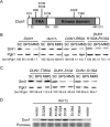
Structure-function analysis of Dun1 protein domains required for Sml1 protein decline in response to iron deficiency. (A) Schematic representation of the most relevant Dun1 domains and amino acid residues. The numbers indicate the amino acid positions. (B) The integrity of the Dun1 FHA domain is essential for the drop in Sml1 protein caused by BPS treatment. Yeast dun1Δ (SPY350) cells transformed with plasmid p413-DUN1(EcoRI) (DUN1), pRS413 (dun1Δ), p413-DUN1-R60A (DUN1-R60A), or p413-DUN1-K100A-R102A (DUN1-K100A-R102A) were grown as described in the legend to Fig. 1. (C) Sml1 protein levels in yeast cells lacking specific Dun1 phosphorylation sites. Yeast dun1Δ (SPY350) cells transformed with plasmid p413-DUN1-T380A (DUN1-T380A), p413-DUN1-S10A (DUN1-S10A), or p413-DUN1-S139A (DUN1-S139A) were grown as described in the legend to Fig. 1. For each transformant in panels B and C, quantitation of Sml1/Pgk1 protein levels under BPS and MMS conditions is relative to the corresponding values obtained in SC medium. A representative image and the average and the standard deviation of three independent biological replicates are shown for each transformant. (D) Mutant Dun1 protein levels under low-iron conditions. Yeast dun1Δ (SPY350) cells transformed with plasmid pRS416, pMH62 (DUN1-D328A) p413-DUN1-R60A (R60A), p413-DUN1-K100A,R102A (K100A-R102A), p413-DUN1-S10A (S10A), p413-DUN1-S139A (S139A), or p413-DUN1-T380A (T380A) were grown at 30°C for 6 h in SC medium with 100 μM BPS. Dun1 protein levels were determined by immunoblotting with an anti-Dun1 antibody, and Ponceau staining was used as a loading control.
FIG 3
FIG 3
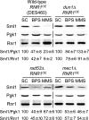
Mec1 and Rad53 checkpoint kinases do not regulate Sml1 protein levels under low-Fe conditions. Wild-type DES460, dun1Δ (MHY307), rad53Δ (DES453), and mec1Δ (DES459) yeast strains overexpressing RNR1 were grown at 30°C for 6 h in SC medium minus tryptophan (SC−trp), SC−trp with 100 μM BPS, and SC−trp with 0.04% MMS added during the last hour. Sml1/Pgk1 and Sml1/Rnr1 protein levels were determined and analyzed as described in the legend to Fig. 1. An anti-Rnr1 antibody was used to determine Rnr1 protein levels. For each strain, Sml1/Pgk1 and Sml1/Rnr1 protein quantitation under BPS and MMS conditions is relative to the values obtained in SC medium. A representative experiment and the average and standard deviation of three independent biological replicates are shown for each strain.
FIG 4
FIG 4
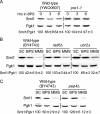
Iron deficiency promotes the 26S proteasomal and vacuolar degradation of Sml1 protein. (A) Wild-type YWO0607 and pre1-1 (YWO0608) yeast strains were grown in SC medium for 3 h at 37°C. Then, 100 μM BPS was added, and the cells were incubated for 6 additional hours at 37°C. (B) Wild-type BY4743, rad6Δ (SPY485), and ubr2Δ (SPY487) yeast strains were grown at 30°C for 6 h in SC medium, SC medium with 100 μM BPS, and SC medium with 0.04% MMS added during the last hour. (C) Wild-type BY4743 and pep4Δ (SPY496) yeast strains were grown at 30°C for 8 h in SC medium, SC medium with 100 μM BPS, and SC medium with 0.04% MMS added during the last hour. In all cases Sml1/Pgk1 protein levels were determined and analyzed as described in the legend to Fig. 1. For each strain, Sml1/Pgk1 protein quantitation under BPS and MMS conditions is relative to the levels obtained in SC medium. A representative experiment and the average and standard deviation of three independent biological replicates are shown.
FIG 5
FIG 5
Identification of genes involved in the regulation of Sml1 protein levels by low iron. (A) Sml1 protein abundance decreases in cells defective in members of the Fe deficiency-sensing pathway. Wild-type W303-1A, Gal-NFS1, Gal-YAH1, Gal-NBP35, Gal-NAR1, and grx3ΔGal-GRX4 yeast strains were grown at 30°C for 40 h in SC medium to repress the expression of the GAL-driven genes. As described in Materials and Methods, the cells were maintained in exponential phase during the whole incubation. (B) Sml1 protein levels in yeast strains with altered Aft1 activity. Wild-type BY4741, aft1Δ, and aft1Δ transformed with plasmid pAFT1-1up were grown at 30°C for 6 h in SC medium or SC medium with 100 μM BPS. (C) FET3 mRNA levels in yeast strains with altered Aft1 activity. Yeast cells were grown as for panel B, total RNA was extracted, and FET3 mRNA levels were determined by quantitative RT-PCR as described in Materials and Methods. FET3 mRNA values were normalized with ACT1 mRNA. (D) Dun1 kinase is required for the Sml1 protein drop observed in Gal-NFS1 cells. Wild-type BY4741, Gal-NFS1 (AXY1988), and dun1ΔGal-NFS1 (AXY2059) cells were grown as described in the legend to panel A. (E) Dun1 kinase is required for the Sml1 protein drop observed in grx3Δ grx4Δ cells. Wild-type BY4741, grx3Δ grx4Δ, and dun1Δ grx3Δ grx4Δ (AXY1926) cells were grown at 30°C in SC medium to exponential growth phase. In panels A, B, D, and E, Sml1/Pgk1 protein levels were determined as described in the legend to Fig. 1 and are relative to the values obtained for the wild-type strain grown in SC medium. In all cases, a representative experiment and the average and standard deviation from at least three independent biological replicates are represented.
FIG 6
FIG 6
Genetic interactions between dun1Δ, sml1Δ, and fet3Δ fet4Δ yeast mutants. Wild-type (BY4741), dun1Δ (SPY350), sml1Δ (SPY589), fet3Δ fet4Δ (SPY386), dun1Δ fet3Δ fet4Δ (AXY1928), sml1Δ fet3Δ fet4Δ (AXY2243), and dun1Δ sml1Δ fet3Δ fet4Δ (AXY2141) yeast strains were grown to exponential phase and either inoculated in liquid SC medium (3 days at 28°C) with the A600 determined every hour with a Spectrostar Nano absorbance microplate reader (A) or spotted in 5-fold serial dilutions on SC medium plates (2 days at 30°C) (B). An experiment representative of at least three independent biological replicates is shown.
FIG 7
FIG 7
Cells lacking DUN1 display defects in dATP and dCTP synthesis during iron scarcity. Wild-type BY4741 and dun1Δ (SPY350) cells were incubated in SC medium or SC medium with 100 μM BPS for 7 h, and dATP and dCTP levels were determined as described in Materials and Methods. The average and standard deviation from three independent biological replicates are represented. The values are relative to the levels obtained for the wild-type strain grown in SC medium. The asterisk indicates statistically significant (P < 0.05) differences between dNTP levels of wild-type cells grown in BPS and the rest of the conditions assayed.
FIG 8
FIG 8
Rnr1 protein levels under iron-deficient conditions. (A) Rnr1 protein levels increase in response to iron deficiency. Wild-type BY4741 and W303-1A strains were grown at 30°C for 6 h in SC medium, SC medium with 100 μM BPS, or SC medium with 0.2 mg/liter 4-NQO added during the last 2 h. (B) Rnr1 protein levels in iron deficiency are not altered in dun1Δ mutant cells. Wild-type (BY4741) and dun1Δ (SPY350) cells were grown as for panel A. (C) Rad53 does not regulate Rnr1 protein levels under iron-deficient conditions. Wild-type (W303-1A), sml1Δ (MHY375), and sml1Δ rad53Δ (MHY380) cells were grown as for panel A. In all cases, Rnr1 protein levels were determined by immunoblotting with an anti-Rnr1 antibody, and Pgk1 protein levels were used as a loading control. An experiment representative of at least three independent biological replicates is shown for each strain.

Similar articles

Cited by

References

    1. Kolberg M, Strand KR, Graff P, Andersson KK. 2004. Structure, function, and mechanism of ribonucleotide reductases. Biochim. Biophys. Acta 1699:1–34. 10.1016/j.bbapap.2004.02.007 - DOI - PubMed
    1. Nordlund P, Reichard P. 2006. Ribonucleotide reductases. Annu. Rev. Biochem. 75:681–706. 10.1146/annurev.biochem.75.103004.142443 - DOI - PubMed
    1. Cotruvo JA, Stubbe J. 2011. Class I ribonucleotide reductases: metallocofactor assembly and repair in vitro and in vivo. Annu. Rev. Biochem. 80:733–767. 10.1146/annurev-biochem-061408-095817 - DOI - PMC - PubMed
    1. Sanvisens N, de Llanos R, Puig S. 2013. Function and regulation of yeast ribonucleotide reductase: cell cycle, genotoxic stress and iron availability. Biomed. J. 36:51–58. 10.4103/2319-4170.110398 - DOI - PubMed
    1. Sun Z, Fay DS, Marini F, Foiani M, Stern DF. 1996. Spk1/Rad53 is regulated by Mec1-dependent protein phosphorylation in DNA replication and damage checkpoint pathways. Genes Dev. 10:395–406. 10.1101/gad.10.4.395 - DOI - PubMed

Publication types

MeSH terms

Substances

LinkOut - more resources