. 2014 Sep;34(17):3341-53.
doi: 10.1128/MCB.00687-14. Epub 2014 Jun 23.

Novel functional complexity of polycystin-1 by GPS cleavage in vivo: role in polycystic kidney disease

Affiliations

Novel functional complexity of polycystin-1 by GPS cleavage in vivo: role in polycystic kidney disease

Almira Kurbegovic et al. Mol Cell Biol. 2014 Sep.

Abstract

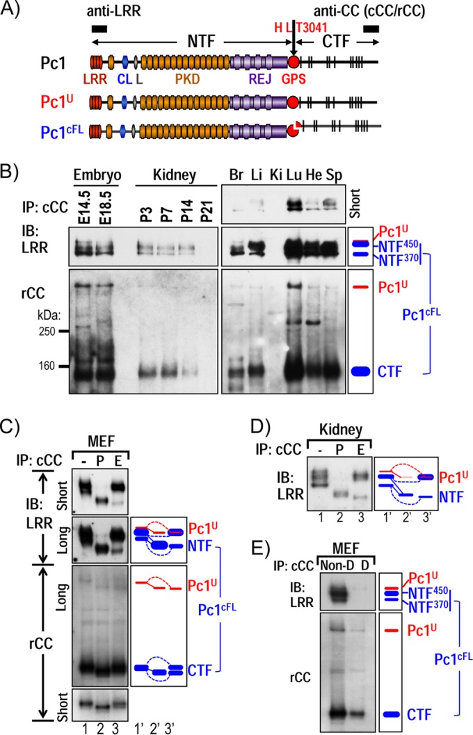
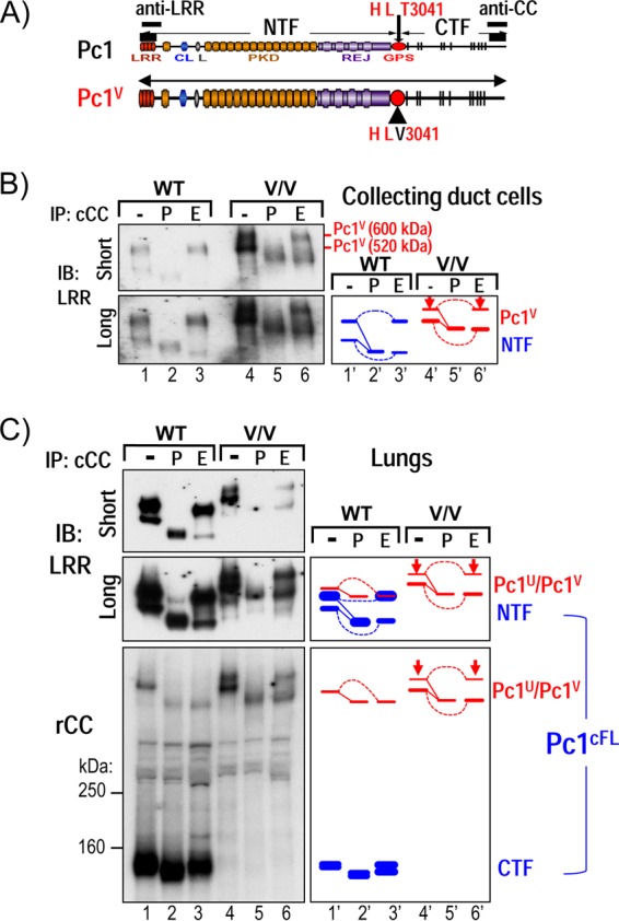
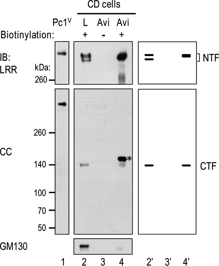
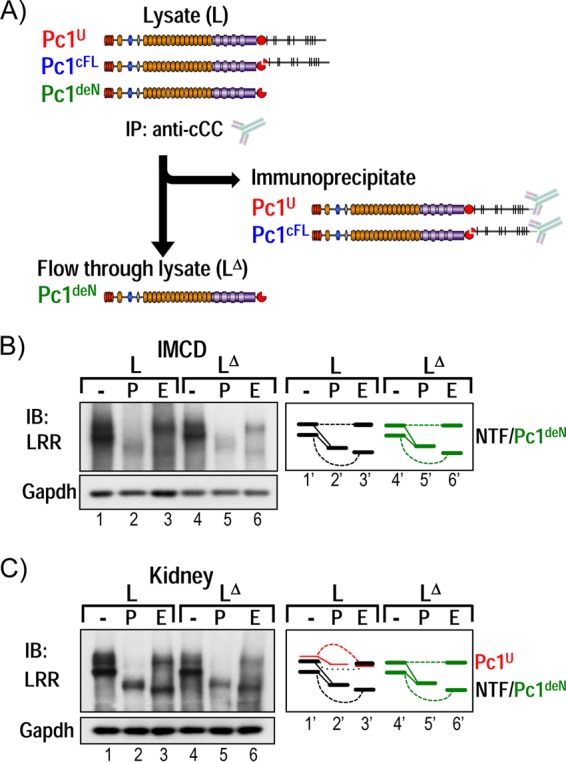
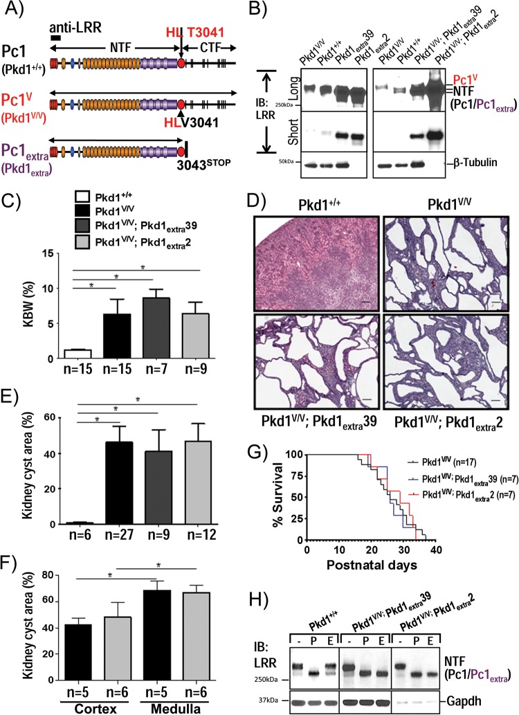
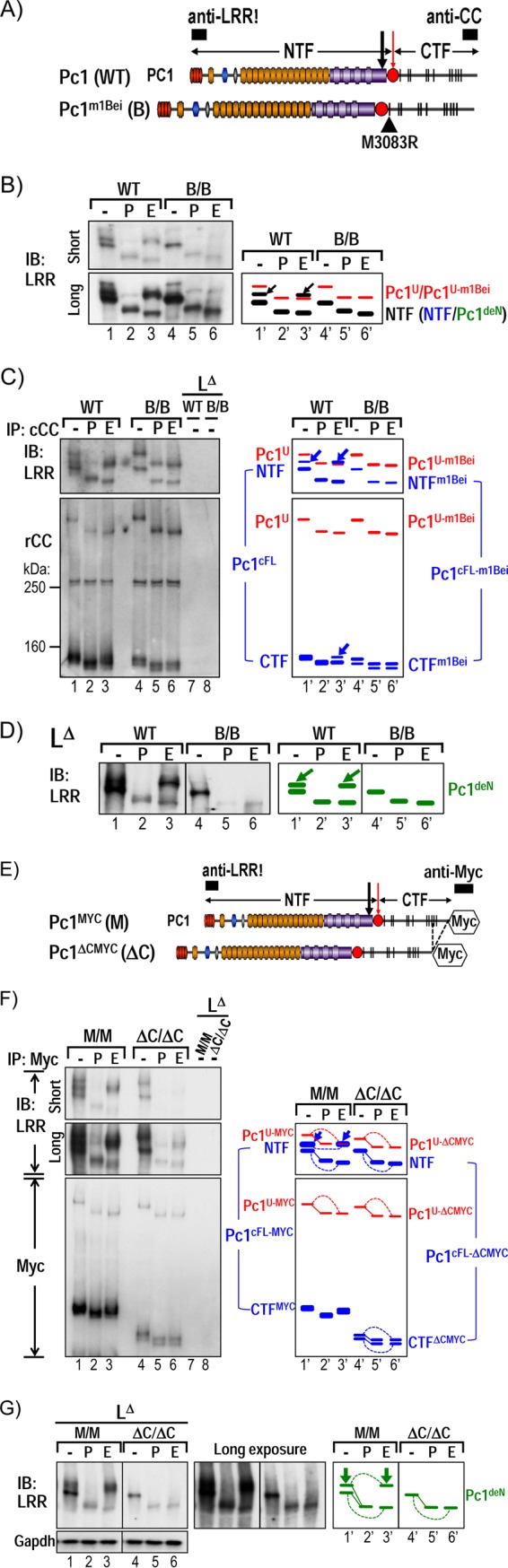
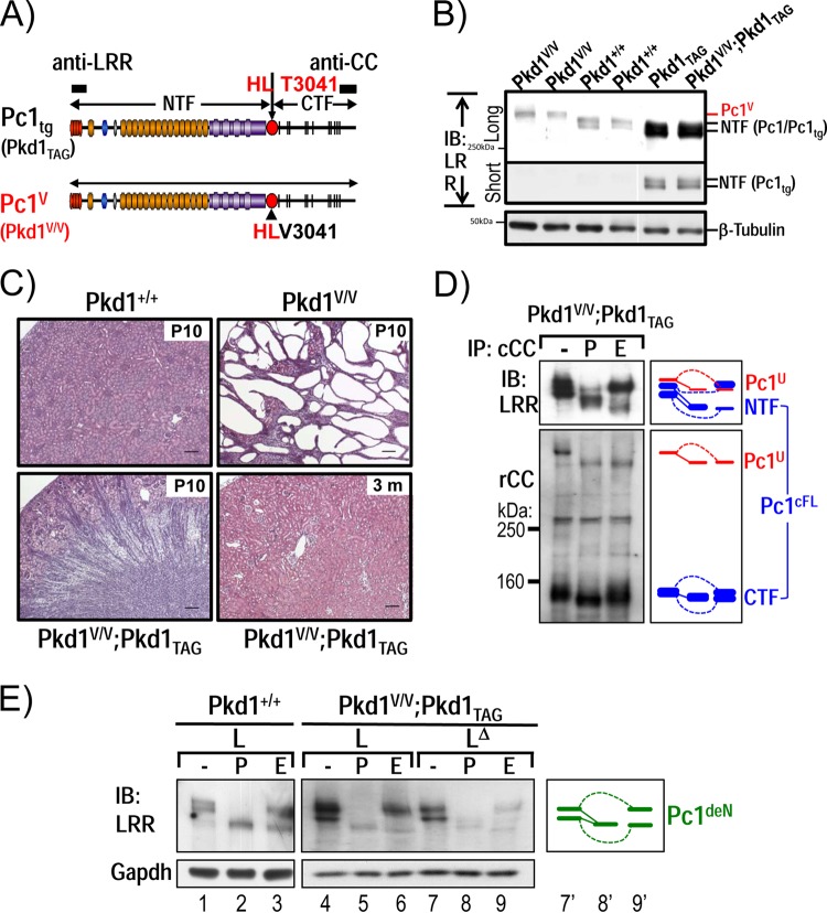
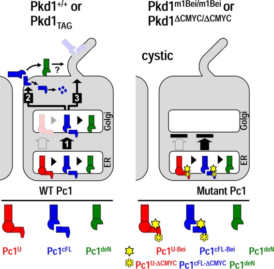
Polycystin-1 (Pc1) cleavage at the G protein-coupled receptor (GPCR) proteolytic site (GPS) is required for normal kidney morphology in humans and mice. We found a complex pattern of endogenous Pc1 forms by GPS cleavage. GPS cleavage generates not only the heterodimeric cleaved full-length Pc1 (Pc1(cFL)) in which the N-terminal fragment (NTF) remains noncovalently associated with the C-terminal fragment (CTF) but also a novel (Pc1) form (Pc1(deN)) in which NTF becomes detached from CTF. Uncleaved Pc1 (Pc1(U)) resides primarily in the endoplasmic reticulum (ER), whereas both Pc1(cFL) and Pc1(deN) traffic through the secretory pathway in vivo. GPS cleavage is not a prerequisite, however, for Pc1 trafficking in vivo. Importantly, Pc1(deN) is predominantly found at the plasma membrane of renal epithelial cells. By functional genetic complementation with five Pkd1 mouse models, we discovered that CTF plays a crucial role in Pc1(deN) trafficking. Our studies support GPS cleavage as a critical regulatory mechanism of Pc1 biogenesis and trafficking for proper kidney development and homeostasis.


Figures

FIG 1
FIG 1
Characterization of endogenous Pc1U and Pc1cFL molecules in normal mouse tissues. (A) Schematic structure of mouse polycystin-1 (Pc1). LRR, leucine-rich repeat; CL, C-type lectin; L, LDL-A; PKD, polycystic kidney disease repeats; REJ, receptor for egg jelly; GPS, G-protein-coupled receptor proteolytic site. Pc1 cleavage occurs at HL↓T3041 site in the GPS motif, resulting in NTF and CTF fragments. Epitope positions of anti-LRR and anti-CC (chicken, cCC; rabbit, rCC) are shown by black boxes. The uncleaved full-length Pc1U (red) and the full-length cleaved Pc1cFL (blue) are schematized. The color code is maintained throughout the figures. (B) Endogenous Pc1 products were analyzed by immunoprecipitation (IP) with anti-cCC from wild-type (WT) mouse embryos (E14.5 and E18.5), kidneys (P3 to P21), and adult (2-month-old) tissues (Br, brain; Li, liver; Ki, kidney; Lu, lung; He, heart; Sp, spleen) and detected by immunoblotting (IB) with anti-LRR (upper panel) and anti-rCC (lower panel). The schematic diagram (right panel) provides an identification guide. (C) N-glycosylation modification of endogenous Pc1 from WT MEFs was monitored by IB on anti-cCC immunoprecipitates, either untreated (−) or treated with PNGase F (P) or endo-H (E). Pc1 products were detected with anti-LRR and anti-rCC from different exposures. The exclusive endo-H sensitivity of Pc1U contrasts with the partial endo-H sensitivity of Pc1cFL. Note that endo-H-deglycosylated Pc1U overlapped with the intense endo-H-resistant NTF450 band (lane 3). A schematic diagram provides an identification guide. (D) N-glycosylation modification of endogenous Pc1 from WT kidneys at P5 was analyzed as described for panel C and detected by IB with anti-LRR. Endogenous Pc1U is endo-H sensitive, whereas the Pc1 NTF subunit is both endo-H resistant and sensitive. (E) Noncovalent association of Pc1cFL subunits. MEF lysates were subjected to IP with anti-cCC under either nondenaturing conditions with 0.5% Triton X-100 (Non-D) or denaturing conditions with detergent SDS (0.1%; D), followed by IB with anti-LRR or anti-rCC. The NTF subunit was coprecipitated by the CTF subunit only under nondenaturing conditions. The schematic diagram provides an identification guide.
FIG 2
FIG 2
GPS cleavage is not a prerequisite for Pc1 intracellular trafficking. (A) Schematic structure of WT Pc1 and noncleavable Pc1V with a T3041V substitution at the HL↓T3041 cleavage consensus site, corresponding to the length of Pc1U. (B) N-glycosylation modification of Pc1 from collecting duct (CD) cells derived from WT and Pkd1V/V (V/V) postnatal kidneys was analyzed by IP with anti-cCC, either untreated (−) or treated with PNGase F (P) or endo-H (E), and then detected by IB with anti-LRR. In Pkd1V/V CD cells, the upper Pc1V band is endo-H resistant (arrow), and the lower Pc1V band is endo-H sensitive, as indicated in the schematic diagram. Note that Pc1U from WT CD cells was not detectable (lane 1). (C) N-glycosylation modification of Pc1 from WT and Pkd1V/V (V/V) postnatal lungs was analyzed for anti-cCC IP products with anti-LRR and anti-rCC, similarly as described for panel B. Of note, Pc1U is weakly detected in the WT lungs, indicated by a red line in the right diagram.
FIG 3
FIG 3
Identification and characterization of Pc1 products on cell surfaces of collecting duct (CD) cells. Confluent CD monolayers were untreated (lane 3) or treated with sulfo-NHS-SS-biotin [sulfosuccinimidyl 2-(biotinamido)-ethyl-1,3-dithiopropionate] (lane 4). Protein lysates were prepared and incubated with NeutrAvidin-agarose (Avi, lanes 3 and 4). The bound proteins were eluted and analyzed by Western blotting using antibodies as indicated. The lack of detection of GM130 (a cis-Golgi protein) in the biotinylated protein population (lane 4) indicates that surface proteins were exclusively biotinylated. Total cell lysate (L) treated with biotin served as a positive control for NTF and CTF (lane 2), with an amount loaded that is equivalent to 1/20 of the amount used for NeutrAvidin-agarose binding. Recombinant Pc1V served to indicate the position of uncleaved Pc1 (lane 1). The schematic diagram at right provides an identification guide. The asterisk indicates a nonspecific band that is seen in the surface protein population (lane 4).
FIG 4
FIG 4
Identification of a novel endogenous Pc1 form: Pc1deN. (A) Immunodepletion strategy to identify the Pc1 NTF detached from the CTF subunit, Pc1deN. Pc1U and Pc1cFL are exhaustively immunoprecipitated from total lysates (L) with anti-cCC, and the putative Pc1deN is analyzed from immunodepleted lysates (LΔ) with anti-LRR. (B) N-glycosylation of endogenous Pc1deN in IMCD cells was analyzed from total lysate (L) and immunodepleted lysates (LΔ) by IB with anti-LRR, after deglycosylation with PNGase F (P) or endo-H (E) or no treatment (−). Note that the Pc1U form was not detectable. The depleted lysate (LΔ) is devoid of Pc1cFL (data not shown). Of interest, Pc1deN was detected in both endo-H-resistant (upper band) and -sensitive (lower band) forms, as schematically depicted at right. GAPDH was used as a loading control. (C) N-glycosylation of endogenous Pc1deN in P5 WT kidney was analyzed from total lysate (L) and immunodepleted lysates (LΔ) as described for IMCD cells in panel B. In total lysate, Pc1cFL overlapped with Pc1deN. The schematic diagram provides an identification guide.
FIG 5
FIG 5
Analysis of Pc1deN functional role by a Pc1extra-BAC transgene in Pkd1V/V mice. (A) Schematic structure of endogenous Pc1 (Pkd1+/+), Pc1V (Pkd1V/V), and Pc1extra (Pkd1extra) proteins. Pc1extra protein was generated by insertion of a termination translation codon in exon 25 of Pkd1 at aa 3043 immediately following the GPS cleavage site. The epitope recognized by anti-LRR is indicated as a black box. (B) Pc1/Pc1V/Pc1extra protein expression levels in P10 kidneys were analyzed by IB with anti-LRR from mice with the genotypes indicated. Protein loading for Pkd1extra2 and Pkd1V/V; Pkd1extra2 mice was decreased by 10-fold (0.9 μg/lane) relative to all other kidney samples (9 μg). Pc1extra exhibits higher expression levels in line Pkd1extra2 than in line Pkd1extra39 and appears in both lines as a single band in comparison to the doublet detected in the wild-type Pc1. β-Tubulin was used as a loading control. (C) Histogram of the kidney weight-to-body weight ratio (KBW) for all genotypes as indicated. The ratios for the Pkd1V/V; Pkd1extra39, Pkd1V/V; Pkd1extra 2, and Pkd1V/V mice at P10 were significantly increased in comparison to the value for WT mice (*, P < 0.0001). n, number of mice. (D) Histopathological analysis (H&E staining) of Pkd1V/V; Pkd1extra kidneys at P10. Pkd1V/V; Pkd1extra39 and Pkd1V/V; Pkd1extra2 mice displayed numerous cysts throughout the kidney parenchyma comparable to Pkd1V/V mice. Scale bar, 100 μm. (E) Histogram of renal cystic index of Pkd1V/V; Pkd1extra kidneys at P10. Cystic involvement (percentage of cystic area) in the Pkd1V/V; Pkd1extra39 and Pkd1V/V; Pkd1extra2 lines shows no significant difference from that in the Pkd1V/V kidneys, but values were highly significant compare to control values (*, P < 0.0001). n, number of mice. (F) Renal cystic involvement in medulla versus cortex in Pkd1V/V and Pkd1V/V; Pkd1extra2 mouse lines at P10. For both Pkd1V/V and Pkd1V/V; Pkd1extra2 mouse lines, cyst surface area (%) is significantly higher in the medulla than in cortex (*, P < 0.0003). Values for the Pkd1V/V; Pkd1extra2 line are not significantly different from those of Pkd1V/V mice in cortex or medulla. n, number of mice. (G) Kaplan-Meier survival curves of the Pkd1V/V, Pkd1V/V; Pkd1extra39, and Pkd1V/V; Pkd1extra2 mice revealed similar life expectancies. (H) Pc1/Pc1V/Pc1extra N-glycosylation status at P10 kidneys was analyzed by IB with anti-LRR on kidney lysates from control Pkd1+/+, Pkd1V/V; Pkd1extra39, and Pkd1V/V; Pkd1extra2 mice, either untreated (−) or deglycosylated with PNGase F (P) or endo-H (E). Pc1 NTF in WT kidneys displayed both Pc1 endo-H-resistant and -sensitive forms, whereas Pc1extra in Pkd1V/V; Pkd1extra39 and Pkd1V/V; Pkd1extra2 kidneys is mainly endo-H sensitive. Protein loading for Pkd1V/V; Pkd1extra2 mice was decreased by 10-fold in comparison to other kidney samples. GAPDH served as a loading control.
FIG 6
FIG 6
Intact CTF is required for intracellular trafficking of the Pc1deN form. (A) Schematic diagram of Pc1 from WT Pkd1 and Pkd1m1Bei alleles. The Pc1m1Bei contains a single substitution (M3083R) in the first TM domain of CTF (black triangle). Epitope positions of anti-LRR and anti-CC are indicated (black boxes). (B) Endogenous Pc1 forms from WT and homozygous Pkd1m1Bei/m1Bei (B/B) embryos (E12.5) were monitored by IB on total lysates either untreated (−) or deglycosylated with PNGase F (P) or endo-H (E) using anti-LRR. Pkd1m1Bei/m1Bei embryos express mutant full-length Pc1U-m1Bei, with exclusive endo-H sensitivity, similar to Pc1U in WT embryos (red). Pc1 NTF in Pkd1m1Bei/m1Bei embryos lacks endo-H resistance relative to WT embryos (arrows). Schematic diagram identifies the corresponding bands. (C) N-glycosylation status of endogenous Pc1U and Pc1cFL forms from WT and mutant Pkd1m1Bei/m1Bei embryos was monitored by IB on anti-cCC immunoprecipitates, either untreated (−) or treated with PNGase F (P) or endo-H (E). Pc1 products were detected with anti-LRR and anti-rCC as indicated. The absence of endo-H resistance of both Pc1 NTF (as observed for total NTF in panel B) and CTF subunits in Pkd1m1Bei/m1Bei embryos contrasts with endo-H resistance in WT embryos (blue, arrows). Re-IP of the flowthrough fractions with anti-cCC (LΔ) confirmed complete depletion of Pc1U and Pc1cFL from both WT and Pkd1m1Bei/m1Bei/ embryo lysates. The schematic diagram depicts corresponding bands. (D) N-glycosylation status of endogenous Pc1deN was analyzed by IB with anti-LRR from depleted lysates (LΔ) of WT and Pkd1m1Bei/m1Bei embryos following deglycosylation. Pc1deN of the Pkd1m1Bei/m1Bei embryos lacks endo-H resistance relative to WT embryos (arrows), as indicated by the schematic diagram at right. (E to G) Results of N-glycosylation analysis for endogenous Pc1 forms from E12.5 Pkd1MYC/MYC knock-in (M/M) and Pkd1ΔCMYC/ΔCMYC knockout (ΔC/ΔC) embryos using the same method as for the Pkd1m1Bei/m1Bei/ (B/B) embryos in panels A to C, except that anti-Myc was used to immunoprecipitate and detect endogenous Myc-tagged Pc1 molecules.
FIG 7
FIG 7
Functional complementation of Pkd1V/V by Pkd1-BAC transgenic mice. (A) Schematic diagram of Pc1tg (Pkd1TAG) and Pc1V (Pkd1V/V). The epitopes recognized by anti-LRR and anti-CC are indicated as black boxes. (B) Protein expression of P10 kidneys from Pkd1+/+, Pkd1TAG, Pkd1V/V, and Pkd1V/V; Pkd1TAG mice were analyzed by IB using anti-LRR. Pc1 expression in Pkd1TAG and Pkd1V/V; Pkd1TAG mice was increased, and Pc1 migrated as a doublet, like endogenous Pc1. β-Tubulin served as a loading control. (C) Kidney histology (H&E staining) of Pkd1+/+, Pkd1V/V, and Pkd1V/V; Pkd1TAG mice. Pkd1V/V; Pkd1TAG mice showed complete rescue of the Pkd1V/V renal phenotype, similar to the WT controls at P10 and 3 months of age. Scale bar, 100 μm. (D) N-glycosylation status of Pc1 from Pkd1V/V; Pkd1TAG P10 kidneys was monitored by IB on anti-cCC immunoprecipitates, either untreated (−) or treated with PNGase F (P) or endo-H (E). Pc1 products were detected with anti-LRR and anti-rCC as indicated. Pc1cFL and Pc1U patterns in Pkd1V/V; Pkd1TAG kidneys are identical to those of the endogenous Pc1 in WT kidneys shown in Fig. 1D. The schematic diagram indicates different Pc1 forms. (E) Pc1 N-glycosylation status of wild-type Pkd1+/+ and Pkd1V/V; Pkd1TAG P10 kidneys was analyzed using total lysate (L) and immunodepleted lysate (LΔ) by IB with anti-LRR following deglycosylation. Pkd1V/V; Pkd1TAG kidneys produce both endo-H-resistant and -sensitive Pc1deN forms as in WT kidneys (left panel). The schematic diagram provides an identification guide.
FIG 8
FIG 8
Model for the role of GPS cleavage in Pc1 biogenesis, trafficking, and functions. In wild-type kidneys (left panel), Pc1U is rapidly converted to Pc1cFL by GPS cleavage in the ER, resulting in small amounts that may exit the ER (open arrow). The resulting Pc1cFL is the main form that exits the ER (1) and traffics to the plasma membrane/cell-cell junctions (2) or other locations, possibly the primary cilium (3). Some of the Pc1cFL in the ER and Golgi compartment undergoes subunit dissociation, producing Pc1deN. The Pc1cFL on the plasma membrane/cell-cell junctions could also dissociate. The released CTF is likely rapidly degraded, as indicated by dots. The Pc1deN remains associated on the membrane, likely through the interaction with other membrane proteins or lipid modification, and accumulates over time (curved arrow). Pc1deN/Pc1cFL probably plays an important role at the cell membrane and/or at the cilium. In Pkd1m1Bei/m1Bei or Pkd1ΔCMYC/ΔCMYC pups (right panel) the mutant Pc1 is unable to exit the ER (black bar), leading to the development of massive cysts despite proper GPS cleavage. Schematized Pc1 forms in wild-type and mutant mice are illustrated below.

Similar articles

Cited by

References

    1. Gabow PA. 1993. Autosomal dominant polycystic kidney disease. N. Engl. J. Med. 329:332–342. 10.1056/NEJM199307293290508 - DOI - PubMed
    1. Ahrabi AK, Jouret F, Marbaix E, Delporte C, Horie S, Mulroy S, Boulter C, Sandford R, Devuyst O. 2010. Glomerular and proximal tubule cysts as early manifestations of Pkd1 deletion. Nephrol. Dial. Transplant. 25:1067–1078. 10.1093/ndt/gfp611 - DOI - PubMed
    1. Lu W, Shen X, Pavlova A, Lakkis M, Ward CJ, Pritchard L, Harris PC, Genest DR, Perez-Atayde AR, Zhou J. 2001. Comparison of Pkd1-targeted mutants reveals that loss of polycystin-1 causes cystogenesis and bone defects. Hum. Mol. Genet. 10:2385–2396. 10.1093/hmg/10.21.2385 - DOI - PubMed
    1. Piontek KB, Huso DL, Grinberg A, Liu L, Bedja D, Zhao H, Gabrielson K, Qian F, Mei C, Westphal H, Germino GG. 2004. A functional floxed allele of Pkd1 that can be conditionally inactivated in vivo. J. Am. Soc. Nephrol. 15:3035–3043. 10.1097/01.ASN.0000144204.01352.86 - DOI - PubMed
    1. Piontek K, Menezes LF, Garcia-Gonzalez MA, Huso DL, Germino GG. 2007. A critical developmental switch defines the kinetics of kidney cyst formation after loss of Pkd1. Nat. Med. 13:1490–1495. 10.1038/nm1675 - DOI - PMC - PubMed

Publication types

MeSH terms

Substances