. 2014 Jun 24:5:4095.
doi: 10.1038/ncomms5095.

Injectable bioadhesive hydrogels with innate antibacterial properties

Affiliations

Injectable bioadhesive hydrogels with innate antibacterial properties

Michael C Giano et al. Nat Commun. .

Abstract

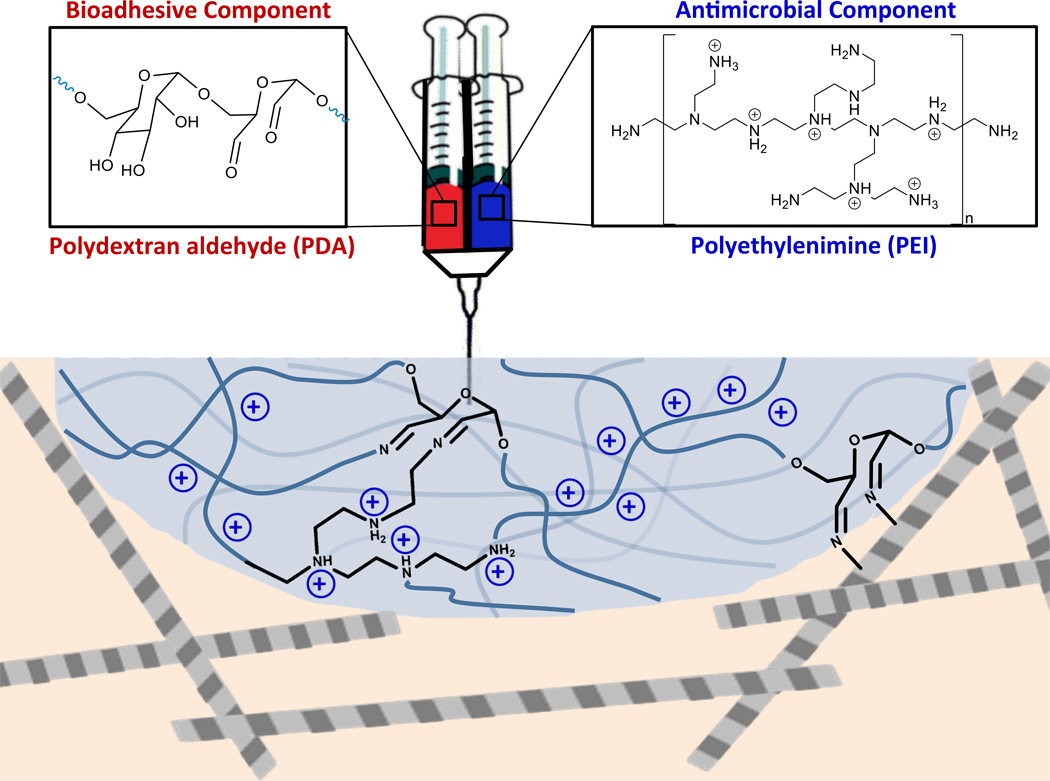
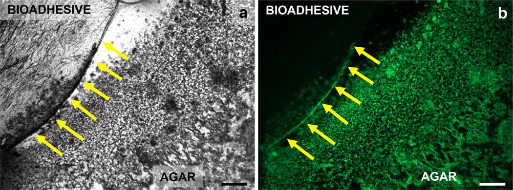
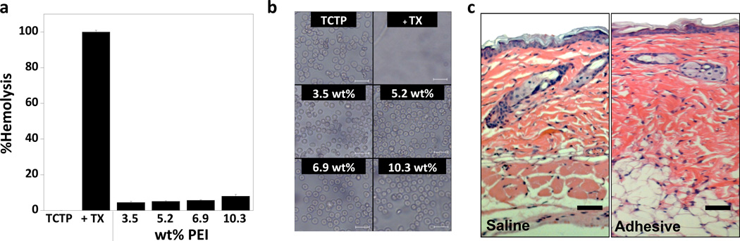
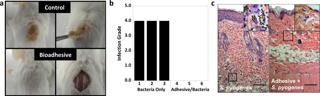
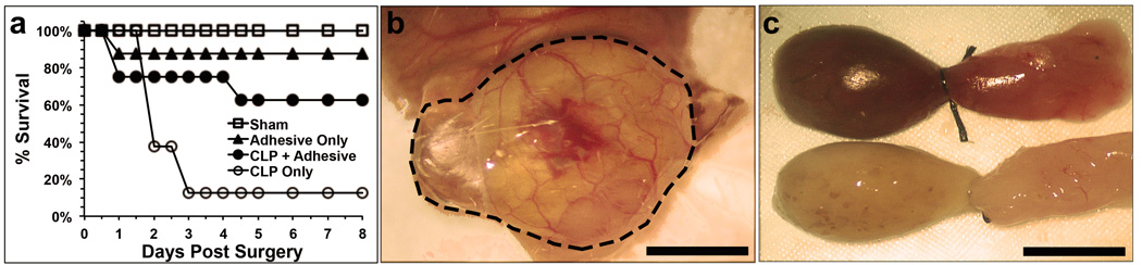
Surgical site infections cause significant postoperative morbidity and increased healthcare costs. Bioadhesives used to fill surgical voids and support wound healing are typically devoid of antibacterial activity. Here we report novel syringe-injectable bioadhesive hydrogels with inherent antibacterial properties prepared from mixing polydextran aldehyde and branched polyethylenimine. These adhesives kill both Gram-negative and Gram-positive bacteria, while sparing human erythrocytes. An optimal composition of 2.5 wt% oxidized dextran and 6.9 wt% polyethylenimine sets within seconds forming a mechanically rigid (~1,700 Pa) gel offering a maximum adhesive stress of ~2.8 kPa. A murine infection model showed that the adhesive is capable of killing Streptococcus pyogenes introduced subcutaneously at the bioadhesive's surface, with minimal inflammatory response. The adhesive was also effective in a cecal ligation and puncture model, preventing sepsis and significantly improving survival. These bioadhesives represent novel, inherently antibacterial materials for wound-filling applications.


Figures

Figure 1
Figure 1. Design of injectable bioadhesive hydrogels with antibacterial activity
The precursor components of the adhesive, polydextran aldehyde (PDA) and polyethylenimine (PEI), define the material’s bioadhesive and antibacterial properties, respectively.
Figure 2
Figure 2. Antibacterial surface properties of adhesive hydrogels
Polydextran aldehyde crosslinked with variable concentrations of PEI were challenged with 104 CFU mL−1 (a) E. coli, or (b) S. aureus, for 24 hours (n = 3). TCTP alone (●) and PDA crosslinked with 6.9 wt% PEI (■) surfaces challenged with increasing concentrations of (c) E. coli, or (d) S. aureus (n = 3; error bars, s.d.).
Figure 3
Figure 3. E. coli viability on antibacterial bioadhesive surfaces
E. coli viability was monitored after 24 hours of incubation on neighboring bioadhesive and agar surfaces (n = 3). Representative images shown. (a) The bright field image defines the boundary between the bioadhesive and agar materials. (b) Viable bacteria fluoresce green only on the agar surface (scale bars = 0.5 mm).
Figure 4
Figure 4. In vitro and in vivo biocompatibility of adhesive
(a) Hemolytic activity of adhesives as a function of PEI wt % along with the control TCTP surface with and without Triton-X (TX) (n = 3; error bars, s.d.). (b) Phase contrast images of hRBCs plated on adhesives of varying PEI wt % or on the control TCTP surface with and without Triton-X (TX) (scale bar = 20 µm). (c) Histological examination of tissue at the injection site for the sham saline injection and the adhesive. No necrosis is evident in either sample and minimal inflammatory response is detected. Cells stained with hematoxylin and eosin, (scale bar = 100 µm). The large region stained purple in the saline image is a hair follicle, while adipose tissue appears white in both images.
Figure 5
Figure 5. In vivo assessment-direct injection of bacteria
(a) Gross external (left) and internal (right) anatomical images of mice injected subcutaneously with 108 CFU mL−1 of S. pyogenes directly into the back (control) or into adhesive (2.5 wt% PDA crosslinked with 6.9 wt% PEI) after 3 days (n = 3). (b) Infection grade of primary tissue at injection site from mice injected with either bacteria only (control) or bacteria injected into adhesive. (c) Histological examination of the inflammatory response to S. pyogenes or adhesive injected with S. pyogenes. Cells stained with hematoxylin and eosin, (scale bar = 100 µm). Insets show 100x magnification of the designated region (scale bar = 5 µm). Inflammatory cells were identified based on cell morphology with →, ⇉, and ► (in yellow) corresponding to neutrophils, macrophages and lymphocytes, respectively.
Figure 6
Figure 6. In vivo assessment-cecal ligation and puncture model
(a) Survival curves for sham, adhesive only, cecal ligation and puncture (CLP) with application of adhesive (2.5 wt% PDA crosslinked with 6.9 wt% PEI), and CLP only (n = 8). (b) Application of adhesive to the punctured cecum during surgery (scale bar = 0.13 cm). Application area outlined. (c) Isolated punctured ceca 24 hours after surgery. Control cecum (top) and experimental cecum that received gel (bottom) are shown (scale bar = 0.25 cm).

Similar articles

Cited by

References

    1. Owens CD, Stoessel K. Surgical site infections: epidemiology, microbiology and prevention. J. Hosp. Infect. 2008;70:3–10. - PubMed
    1. Soper DE, Bump RC, Hurt WG. Wound in infection after abdominal hysterectomy: effect of the depth of subcutaneous tissue. Am. J. Obstet. Gynecol. 1995;173:465–471. - PubMed
    1. Mehdizadeh M, Yang J. Design strategies and applications of tissue bioadhesives. Macromol. Biosci. 2013;13:271–288. - PMC - PubMed
    1. Artzi N, Zeiger A, Boehning F, bon Ramos A, van Vliet K, Edelman ER. Tuning adhesion failure strength for tissue-specific applications. Acta Biomater. 2011;7:67–74. - PMC - PubMed
    1. Lee H, Lee BP, Messersmith PB. A reversible wet/dry adhesive inspired by mussels and geckos. Nature. 2007;448:338-U334. - PubMed

Publication types

MeSH terms

Substances