

Intraobserver variability in fibroid size measurements: estimated effects on assessing fibroid growth
- PMID: 24958408
- PMCID: PMC5452979
- DOI: 10.7863/ultra.33.7.1217
Intraobserver variability in fibroid size measurements: estimated effects on assessing fibroid growth
Abstract
Objectives: To evaluate intraobserver variability of fibroid sonographic measurements and apply this factor to fibroid growth assessment.
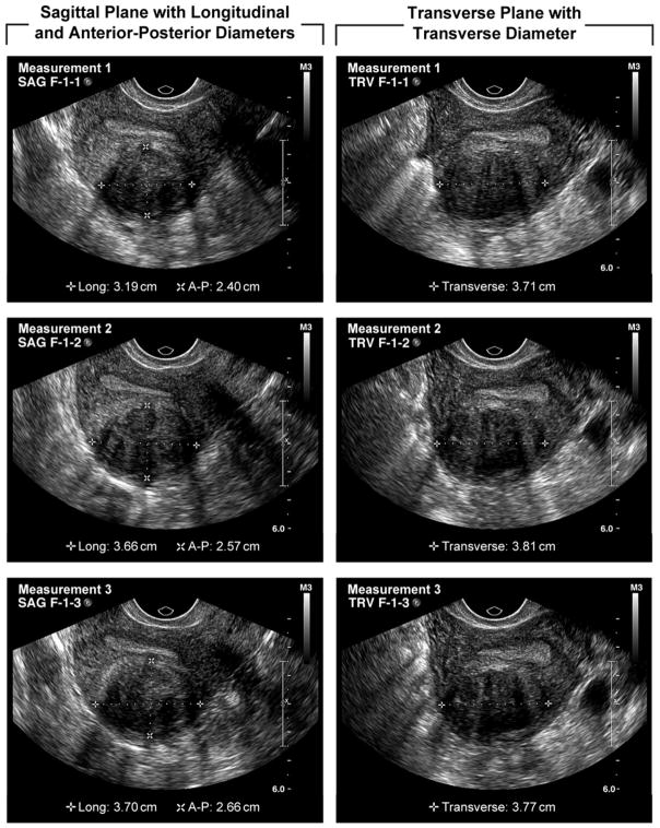
Methods: Study participants were African American women aged 23 to 34 years who had never had a diagnosis of uterine fibroids. All participants underwent transvaginal sonography to screen for the presence of previously undiagnosed fibroids (≥0.5 cm in diameter). The diameters of up to 6 fibroids were measured in 3 perpendicular planes at 3 separate times during the examinations by experienced sonographers. Intraobserver variability as measured by the coefficient of variation (CV) for fibroid diameter and volume was calculated for each fibroid, and factors associated with the CV were assessed by regression models. The impact of variability on growth assessment was determined.
Results: Ninety-six of 300 women screened were found to have at least 1 fibroid, yielding a total of 174 fibroids for this analysis. The mean CV for the 3 measurements of fibroid maximum diameter was 5.9%. The mean CV for fibroid volume was 12.7%. Fibroid size contributed significantly to intraobserver variability (P = .04), with greater variability for smaller fibroids. Fibroid type (submucosal, intramural, or subserosal) was not important. Fibroids from the same woman tended to have similar measurement variability when assessed for volume but not for maximum diameter. Calculations showed that when following up fibroids, as much as a 20% increase in diameter could be due to measurement error, not "true growth."
Conclusions: A small fibroid must have a greater change in size than a large fibroid to conclude that it is growing, but even for small fibroids an increase in diameter of greater than 20% is likely to indicate true growth, not measurement variability.
Keywords: coefficient of variation; fibroid; growth; gynecologic ultrasound; sonography; variability.
© 2014 by the American Institute of Ultrasound in Medicine.
Figures
References
-
- Fedele L, Bianchi S, Dorta M, Zanotti F, Brioschi D, Carinelli S. Transvaginal ultrasonography in the differential diagnosis of adenomyoma versus leiomyoma. Am J Obstet Gynecol. 1992;167:603–6. - PubMed
-
- Kupfer MC, Schiller VL, Hansen GC, Tessler FN. Transvaginal sonographic evaluation of endometrial polyps. J Ultrasound Med. 1994;13:535–9. - PubMed
Publication types
MeSH terms
Grants and funding
LinkOut - more resources
Full Text Sources
Other Literature Sources
Medical
