. 2014 Sep;82(9):3753-63.
doi: 10.1128/IAI.02045-14. Epub 2014 Jun 23.

Intestinal epithelium-specific MyD88 signaling impacts host susceptibility to infectious colitis by promoting protective goblet cell and antimicrobial responses

Affiliations

Intestinal epithelium-specific MyD88 signaling impacts host susceptibility to infectious colitis by promoting protective goblet cell and antimicrobial responses

Ganive Bhinder et al. Infect Immun. 2014 Sep.

Abstract

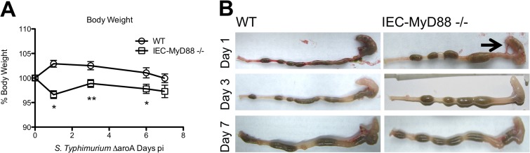
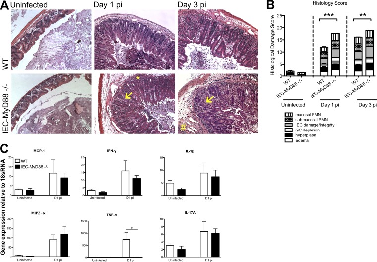
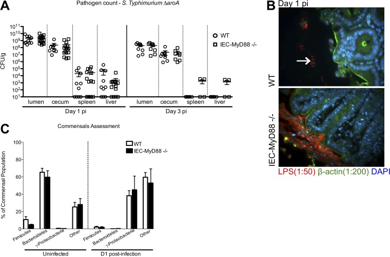
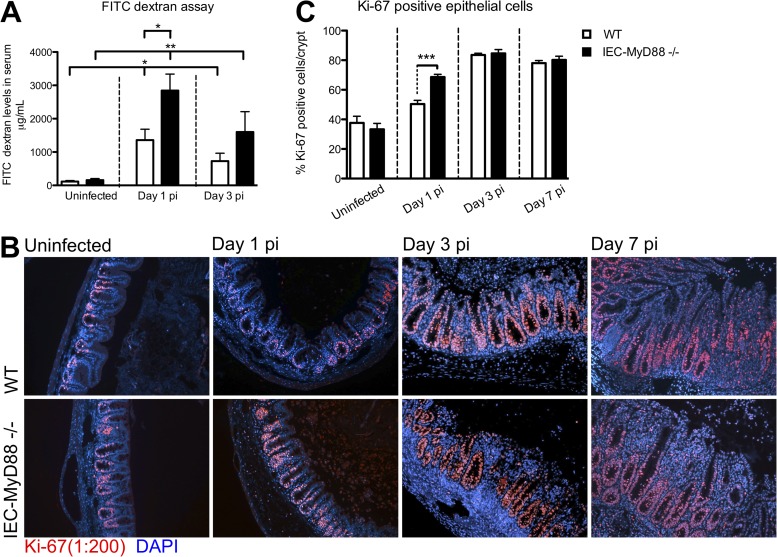
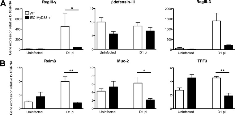
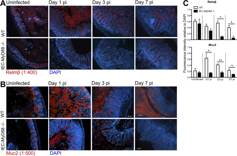
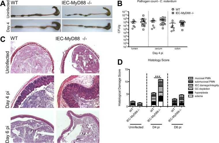
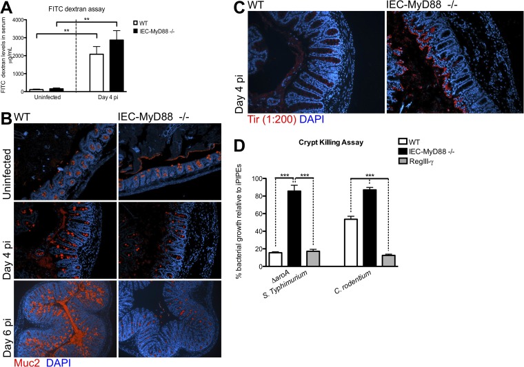
Intestinal epithelial cells (IECs), including secretory goblet cells, form essential physiochemical barriers that separate luminal bacteria from underlying immune cells in the intestinal mucosa. IECs are common targets for enteric bacterial pathogens, with hosts responding to these microbes through innate toll-like receptors that predominantly signal through the MyD88 adaptor protein. In fact, MyD88 signaling confers protection against several enteric bacterial pathogens, including Salmonella enterica serovar Typhimurium and Citrobacter rodentium. Since IECs are considered innately hyporesponsive, it is unclear whether MyD88 signaling within IECs contributes to this protection. We infected mice lacking MyD88 solely in their IECs (IEC-Myd88(-/-)) with S. Typhimurium. Compared to wild-type (WT) mice, infected IEC-Myd88(-/-) mice suffered accelerated tissue damage, exaggerated barrier disruption, and impaired goblet cell responses (Muc2 and RELMβ). Immunostaining revealed S. Typhimurium penetrated the IECs of IEC-Myd88(-/-) mice, unlike in WT mice, where they were sequestered to the lumen. When isolated crypts were assayed for their antimicrobial actions, crypts from IEC-Myd88(-/-) mice were severely impaired in their antimicrobial activity against S. Typhimurium. We also examined whether MyD88 signaling in IECs impacted host defense against C. rodentium, with IEC-Myd88(-/-) mice again suffering exaggerated tissue damage, impaired goblet cell responses, and reduced antimicrobial activity against C. rodentium. These results demonstrate that MyD88 signaling within IECs plays an important protective role at early stages of infection, influencing host susceptibility to infection by controlling the ability of the pathogen to reach and survive at the intestinal mucosal surface.


Figures

FIG 1
FIG 1
IEC-MyD88−/− mice are more susceptible to S. Typhimurium-induced colitis. (A) Body weights of WT and IEC-MyD88−/− mice from D0 to D7 pi, plotted as a percentage of starting weight. IEC-MyD88−/− mice exhibited rapid weight loss by D1 pi that remained significantly below that of WT mice until D6 pi. (B) Unlike WT mice, at D1 pi the IEC-MyD88−/− intestinal tissues displayed severe damage, with severely shrunken ceca devoid of stool contents. The black arrow indicates inflamed ceca, and error bars indicate SEM from at least 6 mice. *, P < 0.05; **, P < 0.005.
FIG 2
FIG 2
IEC-MyD88−/− mice suffer accelerated tissue damage during S. Typhimurium infection. (A) Representative H&E staining of cecal tissues from WT and IEC-MyD88−/− mice taken under uninfected conditions or at D1 and D3 pi. IEC-MyD88−/− mice exhibited increased edema, inflammatory cell infiltrate (*, mucosal infiltration; #, submucosal infiltration), and damage to IEC integrity (arrows) at D1 and D3 pi. (B) Comparative histological damage scores of uninfected and infected (D1 and D3 pi) IEC-MyD88−/− and WT mice. Cecal tissues of IEC-MyD88−/− mice displayed significantly higher histological damage scores at both D1 and D3 pi. Bars represent the damage scores from at least 3 experiments, each with 3 to 5 mice. Error bars indicate SEM. **, P < 0.005; ***, P < 0.0005. (C) Infected WT and IEC-MyD88−/− mice show similar increases in gene transcript levels for the chemokines MCP-1 and MIP2-α, as well as for IFN-γ, IL-1β, and IL-17A. In contrast, compared to WT mice, the IEC-MyD88−/− mice showed significantly decreased transcript levels for TNF-α after infection with the S. Typhimurium ΔaroA mutant. Results are representative of 3 independent infections. Original magnification, ×200.
FIG 3
FIG 3
Disease severity of IEC-MyD88−/− mice is associated with altered S. Typhimurium localization within cecum. (A) No differences in S. Typhimurium ΔaroA mutant pathogen burdens (CFU/g) were identified among the ceca, luminal contents, spleens, or livers from IEC-MyD88−/− and WT mice at D1 or D3 pi. Error bars indicate SEM from at least 9 mice unless otherwise indicated. (B) Immunofluorescence staining for Salmonella LPS (red), β-actin (green), and DNA (blue) in cecal tissues at D1 pi. The presence of MyD88 in IECs (WT) prevents the S. Typhimurium ΔaroA mutant (location indicated by white arrow) from associating closely with the epithelium, sequestering them to the lumen, whereas S. Typhimurium in the IEC-MyD88−/− ceca was found in close proximity to the epithelial surface. Original magnification, ×200. (C) The commensal microbiota found in WT and IEC-MyD88−/− mice under uninfected conditions and after streptomycin treatment at D1 pi, as measured by qPCR. No significant differences were found between the intestinal bacterial populations (at phylum level) present in WT and IEC-MyD88−/− mice.
FIG 4
FIG 4
IEC-MyD88−/− mice display impaired barrier integrity at early infection time points. (A) FITC-dextran-based intestinal permeability assay performed on WT and IEC-MyD88−/− mice under uninfected as well as infected (D1 and D3 pi) conditions. IEC-MyD88−/− mice showed significantly increased barrier permeability compared to that of WT mice at D1 pi. Both groups experienced increased barrier permeability due to infection at D1 and D3 pi compared to that under uninfected conditions. Bars represent the average values for at least 7 mice per group from 3 or more independent experiments. *, P < 0.05; **, P < 0.005. (B) Immunostaining for the proliferation marker Ki-67 (red) and DNA (blue) revealed that WT and IEC-MyD88−/− mice had increased proliferation in cecal tissue beginning at D1 pi. (C) Quantification of percent Ki-67-positive cells per crypt showed there were significantly more proliferating cells in IEC-MyD88−/− mice at D1 pi than in WT mice.
FIG 5
FIG 5
Infection-induced upregulation of gene transcripts for antimicrobial peptides and goblet cell mediators is impaired in IEC-MyD88−/− mice. (A) Infection of WT mice with the S. Typhimurium ΔaroA mutant led to increased gene transcript levels for RegIII-γ (significant) and RegIII-β (not significant) in cecal tissues by D1 pi compared to those of IEC-MyD88−/− mice. (B) IEC-MyD88−/− mice also were impaired in gene transcription levels in cecal tissues for the goblet cell mediators Relmβ, Muc2, and TFF3 at D1 pi compared to levels in WT mice. Error bars represent SEM from three independent experiments (at least 9 mice per group). *, P < 0.05; **, P < 0.005.
FIG 6
FIG 6
Muc2 and Relmβ production is impaired in infected IEC-MyD88−/− mice. (A and B) Representative immunostaining for the goblet cell-specific factors Relmβ (red) (A) and Muc2 (red) (B) in uninfected tissues and D1, D3, and D7 pi cecal tissues, with DNA stained in blue. (C) Fluorescence intensity measurements for Relmβ and Muc2 relative to total DNA staining using ImageJ software revealed WT tissues had significantly greater Relmβ- and Muc2-positive staining at D1, D3, and D7 pi. Bars represent the average fluorescence intensity values, with three measurements per mouse and at least 6 mice per group. *, P < 0.05; **, P < 0.005. Original magnification, ×200.
FIG 7
FIG 7
IEC-MyD88−/− mice suffer accelerated tissue damage during C. rodentium infection. (A) After C. rodentium infection, IEC-MyD88−/− mice displayed shrunken ceca compared to those of WT mice at D4 pi. (B) No differences in C. rodentium tissue pathogen burdens (CFU/g) were identified among the ceca, colons, or luminal contents from IEC-MyD88−/− and WT mice at D4 pi. Error bars indicate SEM from at least 6 mice. (C) Representative H&E staining of cecal tissues from D4 pi WT and IEC-MyD88−/− mice. IEC-MyD88−/− mice exhibit increased edema, hyperplasia, and damage to IEC integrity at D4 pi. (D) Comparative histological damage scores of uninfected and infected (D4 and D6 pi) IEC-MyD88−/− and WT mice. Cecal tissues of IEC-MyD88−/− mice had significantly higher histological damage scores at D4 pi. Bars represent the damage scores of at least 3 experiments, each with 3 to 5 mice. Error bars indicate SEM. ***, P < 0.0005.
FIG 8
FIG 8
IEC-MyD88−/− mice show defects in Muc2 expression and crypt antimicrobial capacity. (A) Both WT and IEC-MyD88−/− mice experience increased barrier permeability at D4 pi. (B) Representative immunostaining for the goblet cell-specific factor Muc2 (red) in uninfected and infected (D4 and D6 pi) cecal tissues, with DNA stained blue. IEC-MyD88−/− mice have decreased positive Muc2 staining at D4 and D6 pi in cecal tissues compared to WT mice. (C) Immunostaining for the C. rodentium translocated effector Tir (red) and DNA (blue) at D4 pi reveals that the lack of MyD88 signaling in IECs allows C. rodentium to more readily infect cecal crypts than in WT mice, where they are sequestered to the lumen. (D) Cecal crypts isolated from WT mice possess greater bactericidal activity against the S. Typhimurium ΔaroA mutant and C. rodentium than crypts from IEC-MyD88−/− mice. Results are plotted as the average growth of bacteria relative to the negative-control iPIPES buffer (100% growth), with 20 μM RegIII-γ incubation presented as a positive control, and are representative of 2 independent experiments with 3 to 5 mice per group.

References

    1. Mandeville KL, Krabshuis J, Ladep NG, Mulder CJ, Quigley EM, Khan SA. 2009. Gastroenterology in developing countries: issues and advances. World J. Gastroenterol. 15:2839–2854. 10.3748/wjg.15.2839 - DOI - PMC - PubMed
    1. Ohl ME, Miller SI. 2001. Salmonella: a model for bacterial pathogenesis. Annu. Rev. Med. 52:259–274. 10.1146/annurev.med.52.1.259 - DOI - PubMed
    1. Akira S, Takeda K. 2004. Toll-like receptor signalling. Nat. Rev. Immunol. 4:499–511. 10.1038/nri1391 - DOI - PubMed
    1. Rakoff-Nahoum S, Paglino J, Eslami-Varzaneh F, Edberg S, Medzhitov R. 2004. Recognition of commensal microflora by Toll-like receptors is required for intestinal homeostasis. Cell 118:229–241. 10.1016/j.cell.2004.07.002 - DOI - PubMed
    1. Gibson DL, Ma C, Bergstrom KSB, Huang JT, Man C, Vallance BA. 2008. MyD88 signalling plays a critical role in host defence by controlling pathogen burden and promoting epithelial cell homeostasis during Citrobacter rodentium-induced colitis. Cell. Microbiol. 10:618–631. 10.1111/j.1462-5822.2007.01071.x - DOI - PubMed

Publication types

MeSH terms

Substances