. 2014 Sep;82(9):3790-801.
doi: 10.1128/IAI.01840-14. Epub 2014 Jun 23.

Protective role of the capsule and impact of serotype 4 switching on Streptococcus mitis

Affiliations

Protective role of the capsule and impact of serotype 4 switching on Streptococcus mitis

Håkon V Rukke et al. Infect Immun. 2014 Sep.

Abstract

The polysaccharide capsule surrounding Streptococcus pneumoniae is essential for virulence. Recently, Streptococcus mitis, a human commensal and a close relative of S. pneumoniae, was also shown to have a capsule. In this study, the S. mitis type strain switched capsule by acquisition of the serotype 4 capsule locus of S. pneumoniae TIGR4, following induction of competence for natural transformation. Comparison of the wild type with the capsule-switching mutant and with a capsule deletion mutant showed that the capsule protected S. mitis against phagocytosis by RAW 264.7 macrophages. This effect was enhanced in the S. mitis strain expressing the S. pneumoniae capsule, which showed, in addition, increased resistance against early clearance in a mouse model of lung infection. Expression of both capsules also favored survival in human blood, and the effect was again more pronounced for the capsule-switching mutant. S. mitis survival in horse blood or in a mouse model of bacteremia was not significantly different between the wild type and the mutant strains. In all models, S. pneumoniae TIGR4 showed higher rates of survival than the S. mitis type strain or the capsule-switching mutant, except in the lung model, in which significant differences between S. pneumoniae TIGR4 and the capsule-switching mutant were not observed. Thus, we identified conditions that showed a protective function for the capsule in S. mitis. Under such conditions, S. mitis resistance to clearance could be enhanced by capsule switching to serotype 4, but it was enhanced to levels lower than those for the virulent strain S. pneumoniae TIGR4.


Figures

FIG 1
FIG 1
Schematic representation of the S. mitis and S. pneumoniae capsule locus and flanking regions. The interspecies homologous regions between S. mitis NCTC 12261T (A) and S. pneumoniae TIGR4 (B) are shown as horizontal brown bars connected by vertical brown lines. The alignment and graphical representation were obtained using the GeVo program, available at https://genomevolution.org/CoGe/GEvo.pl. The dexB gene, shown in yellow, was chosen as the reference gene for alignment. The vertical arrows show the sites of insertion of the erythromycin (Erm) and kanamycin (Km) resistance markers in S. pneumoniae TIGR4, used as the donor of the capsule locus. Black bars, the PCR-amplified regions used to confirm the capsule switch event leading to the insertion of the S. pneumoniae capsule locus into the S. mitis genome. The primer pair used for amplification of fragment a was FP643-FP618, that used for amplification of fragment b was FP617-FP624, and that used for amplification of fragment c was FP623-FP644. (C) Agarose gel electrophoresis imaging of amplicons a, b, and c. Lane M, DNA ladder molecular size marker.
FIG 2
FIG 2
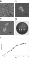
TIGR4 capsule expression by S. mitis. The capsule reaction test (Neufeld test) with pool A antiserum (Statens Serum Institute, Denmark) and the optochin sensitivity test were performed. (A to C) Lack of agglutination of the S. mitis wild type (A), agglutination of the S. mitis TIGR4cps isogenic mutant that acquired the type 4 capsule (B), and agglutination of S. pneumoniae TIGR4 serotype 4 (C). The images in panels A to C were taken with phase-contrast and oil immersion. Bars, 5 μm. (D) Optochin sensitivity test confirming the species identification of (from the left) the S. mitis wild type, S. mitis TIGR4cps, the S. pneumoniae TIGR4 wild type, and S. pneumoniae SP033 (TIGR4 capsule donor). (E) Growth curve of the S. mitis wild type (black circles), S. mitis Δcps (MI016; white circles), and S. mitis TIGR4cps (MI030; black inverted triangles).
FIG 3
FIG 3
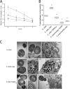
Effect of capsule locus deletion and interspecies capsule switch on S. mitis phagocytosis and intracellular killing by RAW 264.7 macrophages. (A) The S. mitis wild type, S. mitis Δcps, and S. mitis TIGR4cps were incubated with RAW 264.7 macrophages for 1 h to allow phagocytosis. The number of CFU was used to determine intracellular killing over 3 h. The numbers of CFU were normalized to the baseline values determined for the S. mitis wild type. The means and standard deviations from three independent experiments are presented. (B) The wild type and mutants were labeled with FAMSE and then incubated with RAW 264.7 macrophages for 1 h to allow phagocytosis, before the samples were analyzed by flow cytometry. The median proportion of macrophages positive (+ve) for S. mitis is shown (n = 4 or 5). Lines in center of boxes, median values; central boxes, values from the lower to the upper quartiles (25th to 75th percentiles); vertical lines extend from the minimum to the maximum value. **, significantly different from the S. mitis wild type (P < 0.01). (C) TEM images of internalized and extracellular S. mitis and capsule mutants. Bars, 500 nm.
FIG 4
FIG 4
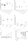
Effect of capsule locus deletion and interspecies capsule switch on S. mitis clearance in mouse models of lung infection and bacteremia and in ex vivo models of blood growth. (A) BALF samples were collected after 4 h of infection. The numbers of CFU ml−1 recovered in BALF are presented. Data were analyzed using one-way ANOVA followed by Dunnett's test using S. mitis as a control. **, P < 0.01; *, P < 0.50. (B and C) Bacterial counts (number of CFU ml−1) in blood of mice challenged intravenously with S. mitis or S. pneumoniae. Inoculum sizes of 5 × 10⁶ CFU ml−1 (B) and 5 × 10⁷ CFU ml−1 (C and D) were used. Blood samples were collected 30 min (C) and 24 h (D) following infection and spread on blood agar plates for determination of viable counts. (A to D) The dot plots show the numbers of CFU obtained for each mouse, with horizontal lines corresponding to median values. (E and F) S. mitis and S. pneumoniae growth and survival in blood. (E) Growth of S. mitis and S. pneumoniae in horse blood. Black symbols, S. mitis strains CCUG12261 (crossed circles), MI016 (circles), MI30.1 (inverted triangles) SK575 (triangles), SK579 (diamonds), and SK597 (squares); white symbols, S. pneumoniae strains TIGR4 (squares), SP035 (inverted triangles), D39 (triangles), and serotype 1 clinical isolate ST306 (circles). (F) S. mitis and S. pneumoniae TIGR4 survival in human blood. The bars are averages from two to three experiments with two to three parallel samples each and represent the fold change in the number of CFU ml−1 after 4 h of incubation in relation to the number of CFU ml−1 in the inoculum. Error bars represent standard errors of the means. Data were analyzed by one-way ANOVA followed by Dunnett's test using S. pneumoniae TIGR4 as a control. *, P < 0.05; **, P < 0.01; a, no colonies were recovered.

Similar articles

Cited by

References

    1. Griffith F. 1928. The significance of pneumococcal types. J. Hyg. (Lond.) 27:113–159. 10.1017/S0022172400031879 - DOI - PMC - PubMed
    1. Bentley SD, Aanensen DM, Mavroidi A, Saunders D, Rabbinowitsch E, Collins M, Donohoe K, Harris D, Murphy L, Quail MA, Samuel G, Skovsted IC, Kaltoft MS, Barrell B, Reeves PR, Parkhill J, Spratt BG. 2006. Genetic analysis of the capsular biosynthetic locus from all 90 pneumococcal serotypes. PLoS Genet. 2:262–269. 10.1371/journal.pgen.0020031 - DOI - PMC - PubMed
    1. Croucher NJ, Harris SR, Fraser C, Quail MA, Burton J, van der Linden M, McGee L, von Gottberg A, Song JH, Ko KS, Pichon B, Baker S, Parry CM, Lambertsen LM, Shahinas D, Pillai DR, Mitchell TJ, Dougan G, Tomasz A, Klugman KP, Parkhill J, Hanage WP, Bentley SD. 2011. Rapid pneumococcal evolution in response to clinical interventions. Science 331:430–434. 10.1126/science.1198545 - DOI - PMC - PubMed
    1. Golubchik T, Brueggemann AB, Street T, Gertz RE, Jr, Spencer CC, Ho T, Giannoulatou E, Link-Gelles R, Harding RM, Beall B, Peto TE, Moore MR, Donnelly P, Crook DW, Bowden R. 2012. Pneumococcal genome sequencing tracks a vaccine escape variant formed through a multi-fragment recombination event. Nat. Genet. 44:352–355. 10.1038/ng.1072 - DOI - PMC - PubMed
    1. Wyres KL, Lambertsen LM, Croucher NJ, McGee L, von Gottberg A, Linares J, Jacobs MR, Kristinsson KG, Beall BW, Klugman KP, Parkhill J, Hakenbeck R, Bentley SD, Brueggemann AB. 2013. Pneumococcal capsular switching: a historical perspective. J. Infect. Dis. 207:439–449. 10.1093/infdis/jis703 - DOI - PMC - PubMed

Publication types

MeSH terms