. 2014 Jun 23;205(6):847-62.
doi: 10.1083/jcb.201312009.

Cleavage by signal peptide peptidase is required for the degradation of selected tail-anchored proteins

Affiliations

Cleavage by signal peptide peptidase is required for the degradation of selected tail-anchored proteins

Jessica M Boname et al. J Cell Biol. .

Abstract

The regulated turnover of endoplasmic reticulum (ER)-resident membrane proteins requires their extraction from the membrane lipid bilayer and subsequent proteasome-mediated degradation. Cleavage within the transmembrane domain provides an attractive mechanism to facilitate protein dislocation but has never been shown for endogenous substrates. To determine whether intramembrane proteolysis, specifically cleavage by the intramembrane-cleaving aspartyl protease signal peptide peptidase (SPP), is involved in this pathway, we generated an SPP-specific somatic cell knockout. In a stable isotope labeling by amino acids in cell culture-based proteomics screen, we identified HO-1 (heme oxygenase-1), the rate-limiting enzyme in the degradation of heme to biliverdin, as a novel SPP substrate. Intramembrane cleavage by catalytically active SPP provided the primary proteolytic step required for the extraction and subsequent proteasome-dependent degradation of HO-1, an ER-resident tail-anchored protein. SPP-mediated proteolysis was not limited to HO-1 but was required for the dislocation and degradation of additional tail-anchored ER-resident proteins. Our study identifies tail-anchored proteins as novel SPP substrates and a specific requirement for SPP-mediated intramembrane cleavage in protein turnover.


Figures

Figure 1.
Figure 1.
A functional proteomics screen in a SPP somatic cell knockout identifies HO-1 as a potential SPP substrate. (A) Odyssey infrared capture of immunoblot for SPP on SPP wild-type (WT, +/+) HCT116, SPP heterozygous (D19 and D12, +/−), or SPP knockout (B4 and B6-1, −/−). (B) Schematic of core/E1 hepatitis C polyprotein expressed from a lentivirus, with signal peptidase (SP) and signal peptide peptidase (SPP) cleavage sites (black arrowheads). The site of the F130E mutation within the amphipathic helix of core is noted (gray arrowhead). The immature (iCore) and mature (mCore) core products, produced after SP and SPP cleavage, are also shown. sig seq, signal sequence. (C) Immunoblot for core antigen in cells expressing WT or mutant (F130E) core in WT (+/+) or SPP knockout (−/−) cells. (D) Immunoblot for mutant (F130E) core antigen in WT (+/+), SPP knockout (−/−), or SPP knockout (−/−) cells transduced with exogenous SPP WT or catalytically inactive SPP D/A mutant, in the presence of mock (top), proteasome (Bortezomib; middle), or the SPP-specific (Z-LL)2 ketone (bottom) inhibitors. (E) Schematic of SILAC mass spectrometry procedure. The experiment was performed in forward and reverse labeling. k/o, knockout; m/z, mass per charge. (F) Results of RNA-Seq analysis for these proteins are also listed. In the reverse SILAC experiment, SPP WT HCT116 cells were cultured in light SILAC media (L), and in the forward experiment, SPP WT HCT116 cells were cultured in heavy SILAC media (H). Reported SILAC ratios in the reverse analysis are H/L, i.e., B4 cells/HCT116 WT cells and, in the forward analysis, are 1/(H/L), i.e., 1/(HCT116 WT cells/B4 cells). Pep represents the unique peptide count. The proteins highlighted in bold type are of type II orientation (either transmembrane or tail anchored). SPP-derived peptides were only identified from SPP HCT116 WT cells and not from SPP knockout B4 cells.
Figure 2.
Figure 2.
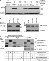
Endogenous HO-1 is degraded in an SPP-dependent pathway. (A–C) Immunoblot for HO-1. (A) WT (HCT116+/+) and SPP knockout (B4−/−) cells were treated with 50 µM hemin (+) for 15 h to induce HO-1. Hemin was removed, and cells were harvested at the times indicated (0/6/12/24 h after hemin induction), using β-actin as a loading control. Cycloheximide was added to all samples after hemin removal to block de novo protein synthesis. (B) WT (HCT116+/+) and SPP knockout (B4−/−) cells were complemented by overexpression of either SPP WT– or SPP D/A–expressing vectors. The complemented cells were harvested 24 h after hemin induction and blotted for HO-1. (C) SPP RNAi– or mock-transfected HeLa cells were treated with 50 µM hemin for 20 h to induce HO-1. Hemin was removed, and cells were harvested at the times indicated after hemin induction (hours postchase) and blotted for HO-1. Cycloheximide was added to all samples after hemin removal to block de novo protein synthesis. Inset shows depletion of SPP at t = 0 h. The chart at the bottom represents the quantification of three independent mock and SPP RNAi transfections. Scanned images were quantified using ImageJ v.1.46r (National Institutes of Health; Schneider et al., 2012). HO-1 levels are normalized against β-actin levels. The error bars represent the standard error.
Figure 3.
Figure 3.
Catalytically active SPP is required for HO-1 degradation. (A) Immunoblot for HO-1. Endogenous HO-1 was induced with hemin (as in Fig. 2 C) in HEK-293T cells, and cells were harvested at indicated times, after incubation with control (DMSO), proteasome inhibitor (lactacystin), or SPP inhibitor ((Z-LL)2 ketone). (B) HA immunoblot of overexpressed HA–HO-1 and HA-Sec11C cotransfected with GFP (mock), WT, or dominant-negative (D/A) SPP. (C) HA and myc immunoblots showing interaction between SPP (D/A) and HO-1. HEK-293T cells were cotransfected with empty myc vector or D265A SPP-myc KKEK (D/A SPP-myc) and either HA–HO-1 or HA-Ube2J1. NP-40/DOC lysates and eluates from the HA and myc immunoprecipitations were probed for the indicated proteins. Approximately 3% of the total cell lysates and ∼25% of the immunoprecipitate eluates were loaded on the gels. Separate blots were exposed independently.
Figure 4.
Figure 4.
HO-1 cleavage by catalytically active SPP is required for its dislocation from the ER membrane and subsequent proteasome-mediated degradation. (A and B) Cleavage of HO-1 was visualized by metabolic label and pulse/chase analysis. Full-length HA–HO-1 (A) or truncated HO-1 (HA–HO-1short; B) plasmids, together with either WT SPP or mutant (D/A) SPP plasmids were cotransfected into HEK-293T cells and [35S]methionine/cysteine pulse labeled for 15 min. HO-1 was recovered by HA immunoprecipitation (IP) at the chase times indicated and analyzed by SDS-PAGE and autoradiography. The black line indicates that the middle lanes from gel were removed, so the second and third lanes were not contiguous. (C) HA–HO-1short was transfected into HEK-293T cells together with either WT or mutant D/A SPP and radiolabeled as in B in the presence or absence of the proteasome inhibitor lactacystin. Postnuclear lysates were separated into membrane fractions (pellet) and soluble fractions (supernatant [Sup]) before HA immunoprecipitation, SDS-PAGE, and autoradiography.
Figure 5.
Figure 5.
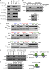
ER-resident tail-anchored proteins are cleaved and degraded in an SPP-dependent pathway. (A) HA immunoblot in HEK-293T cells cotransfected with either HA–HO-1, HA-CYB5A, HA-RAMP4, HA–RAMP4-2, or HA-Ube2J1 and either GFP (mock), WT, or dominant-negative (D/A) SPP as indicated. The SPP inhibitor (Z-LL)2 ketone and the proteasome inhibitor lactacystin (PI) were added to the indicated samples 16 h before harvest. (B) Immunoblot for RAMP4. Mock- or SPP RNAi–transfected HeLa cells were treated with 1 µM tunicamycin for 17 h to induce RAMP4. Cells were harvested at 0 and 24 h after tunicamycin. Unstimulated cells (un.) were harvested at t = 0 h. The inset shows depletion of SPP at t = 0 h. (C) HA immunoblot in HEK-293T cells. Cotransfection with either WT or dominant-negative (D/A) SPP or GFP (−) and full-length or chimeric proteins as indicated. Chimeras contain the cytosolic domain of one protein fused to the transmembrane and luminal domain (TMD) as indicated by the color coding. Black lines indicate that intervening lanes have been spliced out. (D) Immunoblot for GFP in HEK-293T cells transfected with GFP fused to either the TMD of HO-1 (GFP:HO-1TMD) or the TMD of Ube2J1 (GFP:Ube2J1TMD) and either the empty vector (mock), WT, or dominant-negative (D/A) SPP as marked. (E) Immunoblot for GFP in HEK-293T cells transfected with GFP fused to the TMD of HO-1 (GFP:HO-1TMD) and either WT or dominant-negative (D/A) SPP. Samples were treated with the SPP inhibitor (Z-LL)2 ketone or the proteasome inhibitor Bortezomib.
Figure 6.
Figure 6.
The length of the luminal domain is a critical determinant of SPP-mediated cleavage. (A) Immunoblot for HO-1 in HEK-293T cells cotransfected with either N-terminal (HA–HO-1) or C-terminal (HO-1–HA) HO-1 with or without WT, dominant-negative (D/A) SPP, or GFP (mock [M]) as indicated. (B) Immunoblot for GFP in HEK-293T cells transfected with either N-terminal (GFP:HO-1) or C-terminal (HO-1:GFP) GFP-tagged HO-1 with or without WT or dominant-negative (D/A) SPP. The GFP tag used in these constructs contained a double mutation (D82N and F84T; Grotzke et al., 2013) to generate an N-glycosylation site. Samples were subjected to Endoglycosidase H treatment (±Endo H). (C) Immunoblot for HA in HEK-293T cells transfected with HA–HO-1 constructs, which have an additional luminal tag, which starts with (SP) or without (ΔSP) a membrane proximal signal peptidase (SP) cleavage site, followed by a T7 tag and finishing with either a functional C-terminal N-glycosylation site (NIT) or a false N-glycosylation site (QIT) as shown in the schematic. These constructs were cotransfected with GFP (mock; top), WT, or (D/A) SPP and subjected to Endoglycosidase H treatment as indicated.
Figure 7.
Figure 7.
TRC8 ubiquitinates cleaved HO-1. (A) TRC8 expression decreases endogenous HO-1 levels. Immunoblot for HO-1. HEK-293T cells transfected with either GFP (mock), TRC8 FLAG, or TRC8 ΔRING FLAG were induced for 20 h with 50 µM hemin. Cells were harvested at the indicated times after hemin removal. (B) TRC8 ubiquitinates SPP-cleaved HO-1. HA, FLAG, myc, and ubiquitin immunoblots after cotransfection of HA–HO-1 with GFP (mock), TRC8 FLAG, TRC8 ΔRING FLAG, and either SPP myc KEKK (WT) or SPP myc KEKK D265A (D/A), in the presence (+) or absence (−) of Bortezomib. Lysates and HA (HA–HO-1) immunoprecipitations (performed under denaturing conditions) were probed for the indicated proteins.
Figure 8.
Figure 8.
SPP knockout HCT116 cells are competent for US2-mediated MHC class I dislocation. (A) Cytofluorometric analysis of surface MHC class I expression (W6/32) in HCT116 clones transduced with mock lentivirus, US2-expressing lentivirus, or US11-expressing lentivirus. Solid black traces are cells stained with secondary antibody only (anti–mouse Alexa Fluor 647). (B) WT (SPP+/+) and SPP knockout (SPP−/−) HCT116 cells ± HA-US2 were pulse radiolabeled for 10 min, and MHC class I (W6/32) and US2 (rabbit anti-US2) were immunoprecipitated from detergent lysates at the times indicated. IP, immunoprecipitation.

References

    1. Abraham N.G., Kappas A. 2008. Pharmacological and clinical aspects of heme oxygenase. Pharmacol. Rev. 60:79–127 10.1124/pr.107.07104 - DOI - PubMed
    1. Blom D., Hirsch C., Stern P., Tortorella D., Ploegh H.L. 2004. A glycosylated type I membrane protein becomes cytosolic when peptide: N-glycanase is compromised. EMBO J. 23:650–658 10.1038/sj.emboj.7600090 - DOI - PMC - PubMed
    1. Boulant S., Montserret R., Hope R.G., Ratinier M., Targett-Adams P., Lavergne J.P., Penin F., McLauchlan J. 2006. Structural determinants that target the hepatitis C virus core protein to lipid droplets. J. Biol. Chem. 281:22236–22247 10.1074/jbc.M601031200 - DOI - PubMed
    1. Brodsky J.L. 2012. Cleaning up: ER-associated degradation to the rescue. Cell. 151:1163–1167 10.1016/j.cell.2012.11.012 - DOI - PMC - PubMed
    1. Burr M.L., Cano F., Svobodova S., Boyle L.H., Boname J.M., Lehner P.J. 2011. HRD1 and UBE2J1 target misfolded MHC class I heavy chains for endoplasmic reticulum-associated degradation. Proc. Natl. Acad. Sci. USA. 108:2034–2039 10.1073/pnas.1016229108 - DOI - PMC - PubMed

Publication types