. 2014 Jul 15;111(28):10347-52.
doi: 10.1073/pnas.1409233111. Epub 2014 Jun 23.

Piezo1, a mechanically activated ion channel, is required for vascular development in mice

Affiliations

Piezo1, a mechanically activated ion channel, is required for vascular development in mice

Sanjeev S Ranade et al. Proc Natl Acad Sci U S A. .

Abstract

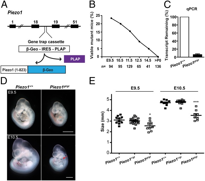
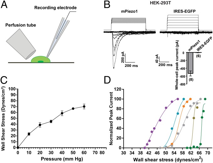
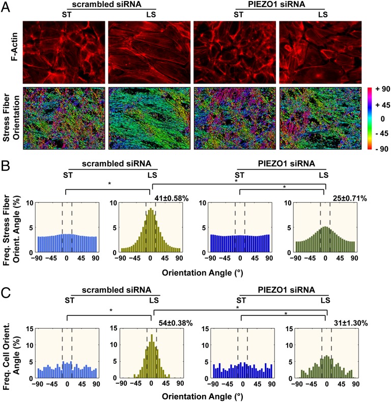
Mechanosensation is perhaps the last sensory modality not understood at the molecular level. Ion channels that sense mechanical force are postulated to play critical roles in a variety of biological processes including sensing touch/pain (somatosensation), sound (hearing), and shear stress (cardiovascular physiology); however, the identity of these ion channels has remained elusive. We previously identified Piezo1 and Piezo2 as mechanically activated cation channels that are expressed in many mechanosensitive cell types. Here, we show that Piezo1 is expressed in endothelial cells of developing blood vessels in mice. Piezo1-deficient embryos die at midgestation with defects in vascular remodeling, a process critically influenced by blood flow. We demonstrate that Piezo1 is activated by shear stress, the major type of mechanical force experienced by endothelial cells in response to blood flow. Furthermore, loss of Piezo1 in endothelial cells leads to deficits in stress fiber and cellular orientation in response to shear stress, linking Piezo1 mechanotransduction to regulation of cell morphology. These findings highlight an essential role of mammalian Piezo1 in vascular development during embryonic development.


Conflict of interest statement

The authors declare no conflict of interest.

Figures

Fig. 1.
Fig. 1.
Piezo1-deficient embryos die at midgestation. (A) Diagram of the Piezo1 genomic locus, gene trap cassette, and the products of Piezo1 gene trap allele (Piezogt ). The exons (black rectangles), β-geo (a fusion between β-gal and neomycin phosphotransferase, blue rectangle), and PLAP (purple rectangle) are indicated. (B) Percentage of viable Piezo1gt/gt mice at different ages derived from Piezo1gt/+ intercrosses. The number of embryos and mice genotyped is shown in the bottom. (C) Relative mRNA expression of Piezo1 in mouse embryonic fibroblasts (MEFs) isolated from E10.5 Piezo1gt/gt embryos compared with Piezo1+/+ MEFs. mRNA levels were determined by quantitative real-time PCR and normalized to glyceraldehyde 3-phosphate dehydrogenase (Gapdh). Bar represents the mean ± SEM (n = 3). (D) Gross appearance of Piezo1gt/gt embryos and their Piezo1+/+ littermates. Red arrow indicates pericardial effusion. (Scale bar: 1 mm.) (E) Size analysis of representative Piezo1+/+, Piezo1+/gt, and Piezo1gt/gt embryos at E9.5 and E10.5. Numbers of embryos used were Piezo1+/+, E9.5 (n = 10), E10.5 (n = 9); Piezo1+/gt, E9.5 (n = 20), E10.5 (n = 13); Piezo1gt/gt, E9.5 (n = 9), E10.5 (n = 8). Bars represent the mean ± SEM *P < 0.05, ***P < 0.001, unpaired two-tailed t test.
Fig. 2.
Fig. 2.
Piezo1 is expressed in embryonic vascular endothelial cells. (A) Representative images of X-gal–stained whole-mount Piezo1+/+ and Piezo+/gt embryos at E9.5 and E10.5. (B) Representative image of E9.5 whole-mount Piezo1+/+ and Piezo1+/gt embryos stained for PLAP and PECAM1 and magnification image of embryonic head showing expression of PLAP and PECAM1 from the same embryos. (C) Representative images of X-gal–stained E11.5 whole-mount hearts and sections of Piezo+/gt embryos. (D) Representative histograms of tdTomato fluorescence in PECAM1+ cells from Piezo1+/+ (gray shaded) or Piezo1+/P1-tdT (red line) embryos. E13.5 embryos and E11.5 embryos were used for heart and yolk sac analysis, respectively, owing to technical consideration for tissue preparation. (E) Histograms of tdTomato fluorescence in PECAM1 cells from embryos in D. Most PECAM1 cells from Piezo1+/tdT yolk sac do not express Piezo1; some PECAM1 cells in Piezo1+/tdT yolk sac also express Piezo1-tdTomato. Stainings in AC were performed in at least four embryos per genotype per time point. Histograms in D and E are representative of at least two embryos per genotype. [Scale bars: 400 μm (A), 500 μm and 200 μm, respectively (B), and 200 μm (C).]
Fig. 3.
Fig. 3.
Piezo1-deficient embryos display vascular remodeling defects. (A) Representative images of PECAM1-stained E8.5 Piezo1+/+ and Piezo1gt/gt whole-mount yolk sacs. (B) Quantification of PECAM1+ area in E8.5 Piezo1+/+ and Piezo1gt/gt whole-mount yolk sac from n = 3 experiments. (C) Representative H&E staining of E9.5 yolk sac cross-sections from Piezo1+/+ and Piezo1gt/gt embryos. (D) Quantification of vessel thickness from H&E staining of E9.5 yolk sac from n = 2 experiments. *P < 0.05, unpaired two-tailed Student t test. [Scale bars: 100 μm (A) and 200 μm (C).]
Fig. 4.
Fig. 4.
Piezo1 is activated by fluid shear stress. (A) Pictorial representation of perfusion tube setup. Whole-cell currents were recorded from HEK293T cells while the cell was stimulated by shear stress induced by directing pulses of fluid flow through a perfusion tube placed relative to the cell. The flow rate is regulated by varying the pressure applied to the fluid inside the tube. (B) Representative traces of whole-cell currents recorded at −80 mV from HEK293T cells expressing mPiezo1 (Left) or IRES-eGFP (Right) in response to 600-ms pulses of increasing laminar shear stress. The stimulus trace above the current indicates the pressure pulse applied to the solution in the perfusion tube (which was later back-calculated to the applied shear stress intensities) and corresponds to the whole-cell current recorded for that stimulus. (Inset) Shear stress-induced average maximum whole-cell peak currents from mPiezo1 (n = 8) and IRES-EGFP (n = 6) transfected cells. Bars indicate mean ± SEM. (C) Pressure–wall shear stress relationship. Particle image velocimetry was used to estimate the wall shear stress intensity at the indicated tube pressure. Each data point is the mean ± SEM from values of five or six independent trials. The measurement was done for the range of pressure (0–60 mmHg) used to activate the cells during the experiment. (D) Wall shear stress to normalized peak current relationship plotted for six individual cells expressing mPiezo1, depicting range of activation. For each cell, currents were normalized to the peak current recorded at maximum shear stress, before losing the seal. The stimulus–response curve was fit with a Boltzmann function to determine the half-maximal response.
Fig. 5.
Fig. 5.
Knockdown of PIEZO1 leads to defects in cellular alignment upon laminar shear stress. (A) F-actin images stained with TRITC-phalloidin (Upper) and angles of stress fiber orientation (Lower). Angle measurements were assigned a color code (right) where 0° is parallel to the flow chamber and −90° and 90° are perpendicular. (B) Histograms of stress fiber orientation of cells. (C) Histograms of cell orientation. The mean ± SEM values refer to percentage of orientations between ±15° for three independent biological experiments. Total numbers of cells analyzed are 348 (ST) and 236 (LS) for scrambled siRNA and 290 (ST) and 170 (LS) for PIEZO1 siRNA. ST, static; LS, laminar shear for 24 h. Asterisk indicates significant difference by Watson’s U2 test.

References

    1. Hoffman BD, Grashoff C, Schwartz MA. Dynamic molecular processes mediate cellular mechanotransduction. Nature. 2011;475(7356):316–323. - PMC - PubMed
    1. Gillespie PG, Müller U. Mechanotransduction by hair cells: Models, molecules, and mechanisms. Cell. 2009;139(1):33–44. - PMC - PubMed
    1. Tsunozaki M, Bautista DM. Mammalian somatosensory mechanotransduction. Curr Opin Neurobiol. 2009;19(4):362–369. - PMC - PubMed
    1. Orr AW, Helmke BP, Blackman BR, Schwartz MA. Mechanisms of mechanotransduction. Dev Cell. 2006;10(1):11–20. - PubMed
    1. Jaalouk DE, Lammerding J. Mechanotransduction gone awry. Nat Rev Mol Cell Biol. 2009;10(1):63–73. - PMC - PubMed

Publication types