. 2014 Jul 8;111(27):9899-904.
doi: 10.1073/pnas.1402238111. Epub 2014 Jun 23.

C/EBPα is an essential collaborator in Hoxa9/Meis1-mediated leukemogenesis

Affiliations

C/EBPα is an essential collaborator in Hoxa9/Meis1-mediated leukemogenesis

Cailin Collins et al. Proc Natl Acad Sci U S A. .

Abstract

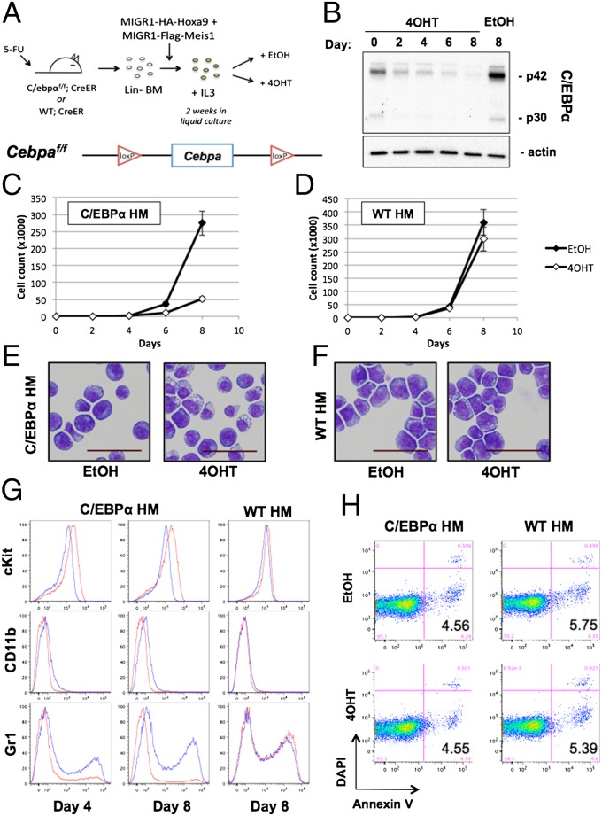
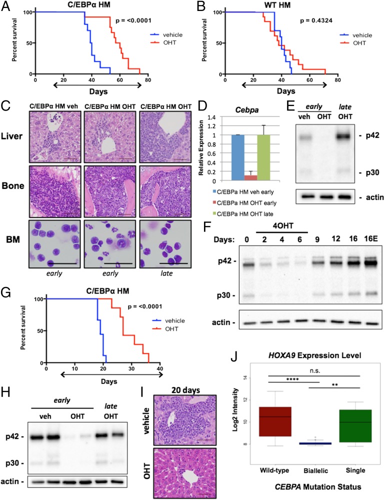
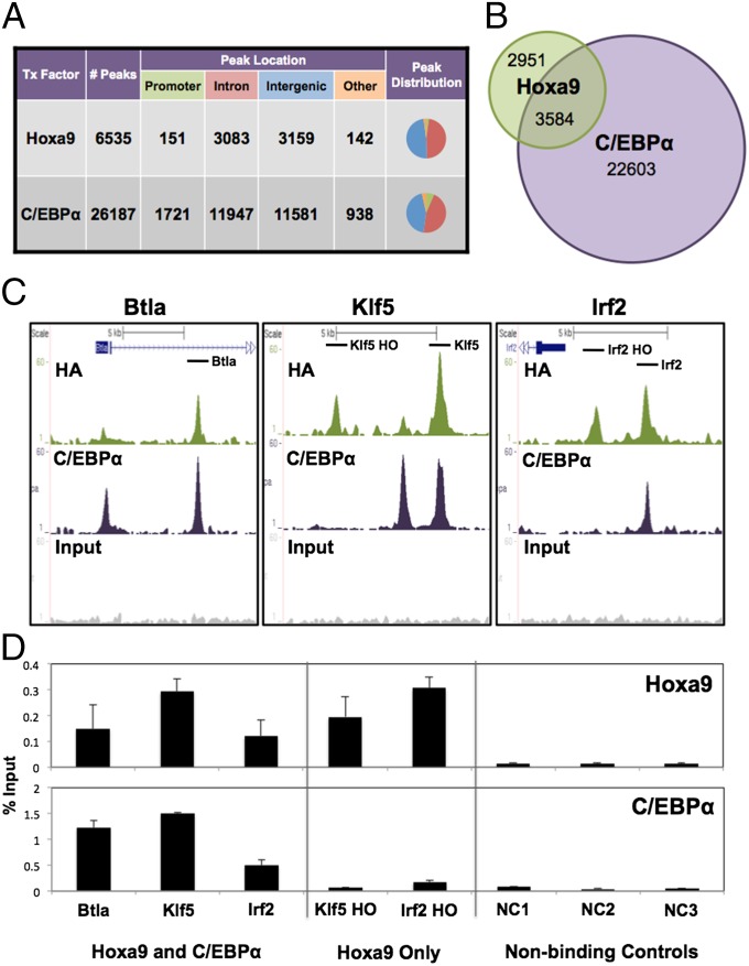
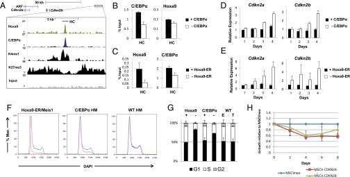
Homeobox A9 (HOXA9) is a homeodomain-containing transcription factor that plays a key role in hematopoietic stem cell expansion and is commonly deregulated in human acute leukemias. A variety of upstream genetic alterations in acute myeloid leukemia (AML) lead to overexpression of HOXA9, almost always in association with overexpression of its cofactor meis homeobox 1 (MEIS1) . A wide range of data suggests that HOXA9 and MEIS1 play a synergistic causative role in AML, although the molecular mechanisms leading to transformation by HOXA9 and MEIS1 remain elusive. In this study, we identify CCAAT/enhancer binding protein alpha (C/EBPα) as a critical collaborator required for Hoxa9/Meis1-mediated leukemogenesis. We show that C/EBPα is required for the proliferation of Hoxa9/Meis1-transformed cells in culture and that loss of C/EBPα greatly improves survival in both primary and secondary murine models of Hoxa9/Meis1-induced leukemia. Over 50% of Hoxa9 genome-wide binding sites are cobound by C/EBPα, which coregulates a number of downstream target genes involved in the regulation of cell proliferation and differentiation. Finally, we show that Hoxa9 represses the locus of the cyclin-dependent kinase inhibitors Cdkn2a/b in concert with C/EBPα to overcome a block in G1 cell cycle progression. Together, our results suggest a previously unidentified role for C/EBPα in maintaining the proliferation required for Hoxa9/Meis1-mediated leukemogenesis.

Keywords: enhancer; gene regulation.


Conflict of interest statement

The authors declare no conflict of interest.

Figures

Fig. 1.
Fig. 1.
C/EBPα is required for Hoxa9/Meis1-mediated transformation. (A) Schematic of cell line generation and Cebpa-targeted allele. 5-FU, 5-flurouracil. (B) C/EBPα HM and WT HM cells were treated for an 8-d time course with 5 nM 4OHT or EtOH, and protein levels in the C/EBPα HM cells were assessed using Western blotting. Cellular proliferation of both C/EBPα HM (C) and WT HM (D) cells was determined by cell counting; data represent mean ± SD of two independent experiments. Cell morphology of C/EBPα HM (E) and WT HM (F) cells was assessed after 8 d. (Scale bars: 50 μm.) (G) Surface expression of c-Kit, CD11b, and Gr1 at days 4 and 8 in C/EBPα HM cells (Left and Center) and WT HM cells (Right) after continuous treatment with 4OHT (red) or EtOH (blue). (H) Annexin V and DAPI staining for apoptotic cells at day 8 of treatment with 4OHT or EtOH. Black numbers in the lower right corner represent the mean number of apoptotic cells (AnnexinV+/DAPI) from four independent experiments. Mean ± SD values are as follows: C/EBPα HM EtOH, 4.56 ± 3.62; C/EBPα HM 4OHT, 4.54 ± 1.23; WT EtOH, 5.74 ± 5.31, and WT 4OHT, 5.38 ± 3.75. Flow cytometry plots from one representative experiment are shown.
Fig. 2.
Fig. 2.
Loss of C/EBPα impairs Hoxa9-mediated leukemogenesis. Survival curves for mice transplanted with C/EBPα HM cells [A; n = 10(veh), 12(OHT); P < 0.0001 by log rank] or WT HM cells [B; n = 10(veh), 14(OHT); P = 0.4324 by log rank]. The treatment period with OHT (red) or vehicle (blue) is indicated by the arrow below the graphs. (C) Tissue histology of liver and bone, and bone marrow (BM) cytospins for C/EBPα HM vehicle- and OHT-treated mice that died before 40 d (Left and Center; “early”) and an OHT-treated mouse that died at 60 d posttransplantation (Right; “late”). (Scale bars: 50 μm.) (D and E) RT-PCR expression of Cebpa and Western blot analysis of C/EBPα protein levels corresponding to samples shown in C (mean ± SD). veh, vehicle. (F) C/EBPα protein levels in cells treated with 4OHT for 6 d and subsequently maintained in the absence of 4OHT for an additional 10 d. The rightmost lane (16E) corresponds to cells treated continuously with EtOH for 16 d. (G) Survival curve of mice transplanted with primary leukemic spleen cells from a C/EBPα HM vehicle-treated mouse [n = 9(veh), 7(OHT); P < 0.0001 by log rank]. The treatment period with OHT (red) or vehicle (blue) is indicated by the arrow below the graph. (H) C/EBPα protein levels in vehicle-treated leukemic mice (Left) compared with OHT-treated mice preleukemic controls (Center) and leukemic OHT-treated mice (Right). (I) Liver histology of leukemic vehicle- and preleukemic OHT-treated mice at 20 d. (Scale bars: 50 μm.) (J) HOXA9 expression level in a cohort of patients with AML subdivided by CEBPA mutation status (n = 344) ****P < 0.0005; **P < 0.005; ns, not sigificant.
Fig. 3.
Fig. 3.
C/EBPα colocalizes with Hoxa9 at promoter distal enhancers. (A) Peak number and distribution of Hoxa9 and C/EBPα ChIP-seq in a Hoxa9/Meis1-transformed cell line (“Other” category contains 5′/3′ UTR and exons). Tx, transcription. (B) Peak overlap between Hoxa9 and C/EBPα ChIP-seq. (C) Representative Hoxa9 (HA)/C/EBPα-cobound loci. Bars indicate location of quantitative PCR (qPCR) primer pairs. (D) Independent ChIP-qPCR validation of ChIP-seq data for Hoxa9 (Upper) and C/EBPα (Lower) binding at Hoxa9–C/EBPα-cobound sites, Hoxa9 only, and nonbinding controls; bars indicate mean ± SD of at least two independent experiments. Btla, B and T lymphocyte associated; Klf5, Kruppel-like factor 5; Irf2, interferon regulatory factor 2.
Fig. 4.
Fig. 4.
C/EBPα and Hoxa9 coregulate expression of Cdkn2a/b. (A) ChIP-seq tracks for Hoxa9, C/EBPα, H3K4me1, and H3K27me3 at the Hoxa9–C/EBPα binding site 50 kb downstream of the Cdkn2a/b locus. Bars indicate the location of the qPCR primer set. HC, Hoxa9/C/EBPα cobound site. ChIP-qPCR for C/EBPα and Hoxa9 binding at the HC binding site after 3-d loss of C/EBPα (B) or Hoxa9 (C). RT-PCR expression of Cdkn2a and Cdkn2b over a 4-d time course after loss of C/EBPα (D) or loss of Hoxa9 (E). (F) Cell cycle analysis at day 6 after loss of Hoxa9 (Left) or C/EBPα (Center), or in EtOH- or 4OHT-treated WT HM cells (Right) (blue, control; red, loss of Hoxa9 or C/EBPα). (G) Quantification of cell cycle profiles analyzed using FlowJo (TreeStar). E, EtOH; T, 4OHT. (H) Relative cellular proliferation of Hoxa9/Meis1-transformed cells coexpressing MSCVneo, MSCV-CDKN2A, or MSCV-CDKN2B. All data are expressed as mean ± SD of at least two independent experiments.

References

    1. Krumlauf R. Hox genes in vertebrate development. Cell. 1994;78(2):191–201. - PubMed
    1. Eklund E. The role of Hox proteins in leukemogenesis: Insights into key regulatory events in hematopoiesis. Crit Rev Oncog. 2011;16(1-2):65–76. - PMC - PubMed
    1. Sitwala KV, Dandekar MN, Hess JL. HOX proteins and leukemia. Int J Clin Exp Pathol. 2008;1(6):461–474. - PMC - PubMed
    1. Golub TR, et al. Molecular classification of cancer: Class discovery and class prediction by gene expression monitoring. Science. 1999;286(5439):531–537. - PubMed
    1. Shah N, Sukumar S. The Hox genes and their roles in oncogenesis. Nat Rev Cancer. 2010;10(5):361–371. - PubMed

Publication types

MeSH terms

Substances

Associated data