. 2014 Jul 8;111(27):9834-9.
doi: 10.1073/pnas.1407898111. Epub 2014 Jun 23.

STAT1-induced ASPP2 transcription identifies a link between neuroinflammation, cell polarity, and tumor suppression

Affiliations

STAT1-induced ASPP2 transcription identifies a link between neuroinflammation, cell polarity, and tumor suppression

Casmir Turnquist et al. Proc Natl Acad Sci U S A. .

Abstract

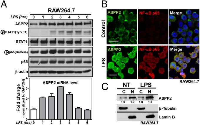
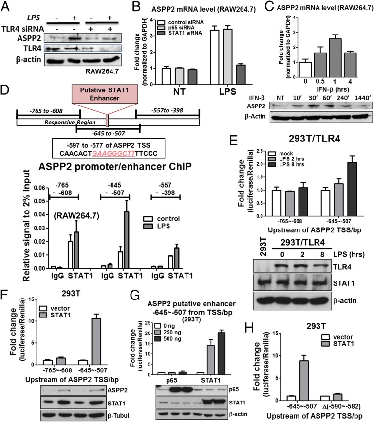
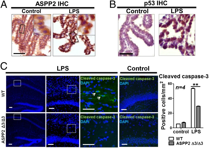
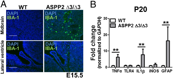
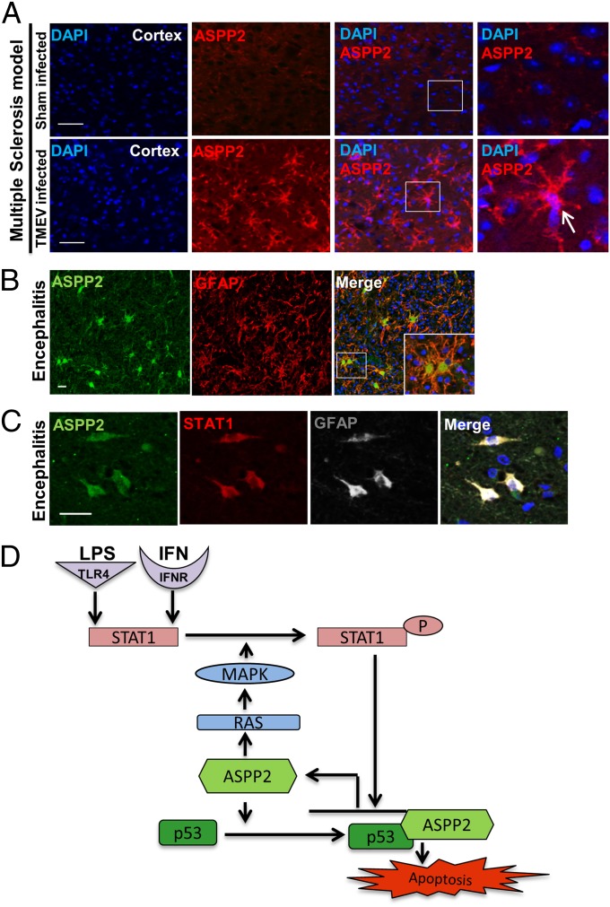
Inflammation and loss of cell polarity play pivotal roles in neurodegeneration and cancer. A central question in both diseases is how the loss of cell polarity is sensed by cell death machinery. Here, we identify apoptosis-stimulating protein of p53 with signature sequences of ankyrin repeat-, SH3 domain-, and proline-rich region-containing protein 2 (ASPP2), a haploinsufficient tumor suppressor, activator of p53, and regulator of cell polarity, as a transcriptional target of signal transducer and activator of transcription 1 (STAT1). LPS induces ASPP2 expression in murine macrophage and microglial cell lines, a human monocyte cell line, and primary human astrocytes in vitro. LPS and IFNs induce ASPP2 transcription through an NF-κB RELA/p65-independent but STAT1-dependent pathway. In an LPS-induced maternal inflammation mouse model, LPS induces nuclear ASPP2 in vivo at the blood-cerebral spinal fluid barrier (the brain's barrier to inflammation), and ASPP2 mediates LPS-induced apoptosis. Consistent with the role of ASPP2 as a gatekeeper to inflammation, ASPP2-deficient brains possess enhanced neuroinflammation. Elevated ASPP2 expression is also observed in mouse models and human neuroinflammatory disease tissue, where ASPP2 was detected in GFAP-expressing reactive astrocytes that coexpress STAT1. Because the ability of ASPP2 to maintain cellular polarity is vital to CNS development, our findings suggest that the identified STAT1/ASPP2 pathway may connect tumor suppression and cell polarity to neuroinflammation.

Keywords: TLR4; TP53BP2; multiple sclerosis.


Conflict of interest statement

The authors declare no conflict of interest.

Figures

Fig. 1.
Fig. 1.
ASPP2 is induced by LPS. (A) LPS time course showing increased ASPP2 expression at protein and mRNA levels in RAW264.7. Expression levels of signaling pathways downstream of LPS were examined, including STAT1 and p65. (B) IF staining of ASPP2 and p65 after 2 h of LPS treatment in RAW264.7. (Scale bars: 10 µm.) (C) Nuclear (N) and cytoplasmic (C) fractionations of ASPP2 on LPS treatment in RAW264.7. Quantification was performed using densitometry analysis. NT, no treatment.
Fig. 2.
Fig. 2.
ASPP2 is a target of STAT1. (A) TLR4 siRNA reduces ASPP2 induction after LPS treatment in RAW264.7 cells. (B) STAT1 siRNA but not p65 siRNA reduces ASPP2 induction after LPS treatment in RAW264.7 cells. (C) IFN-β time course showing increased ASPP2 expression at protein and mRNA levels in RAW264.7. (D) Illustration of the LPS-responsive region of the ASPP2 enhancer region. Putative STAT1 binding site is within the −645 to −507 region. Results of STAT1 ChIP in RAW264.7. The regions corresponding to each of the primer sets are shown. Results are the average of duplicate treatments, and error bars show the range of the duplicates. (E) ASPP2 (−645 to −507) -Luc shows increased activity after LPS treatment for 8 h, whereas ASPP2 (−765 to −608) -Luc shows no response. (F) Only ASPP2 (−645 to −507) -Luc is activated after STAT1 exogenous expression. ASPP2 (−765 to −608) -Luc remains unresponsive. (G) STAT1 but not p65 is able to induce ASPP2 (−645 to −507) -Luc activity. (H) After deletion of the STAT1 binding sequence located at −590 to −582, ASPP2 (−645 to −507) -Luc activity after STAT1 exogenous expression is abolished. NT, no treatment.
Fig. 3.
Fig. 3.
LPS induces nuclear ASPP2 expression in a model of maternal inflammation, and ASPP2 mediates apoptosis. (A) On LPS injection, ASPP2 is disrupted from the TJs and relocalized to the nucleus of CP epithelial cells. (Scale bar: 25 µm.) (B) After LPS injection, p53 appears in the nucleus of CP epithelial cells. (Scale bar: 25 µm.) (C) IF staining of cleaved caspse-3 in LPS-injected and control saline-injected WT and ASPP2 Δ3/Δ3 mice. Arrows indicate cleaved caspase-3–positive cells. (Scale bars: 25 µm.) Quantification of the number of cleaved caspase-3–positive cells in the hippocampus after LPS or saline injection (n = 4). **P < 0.01.
Fig. 4.
Fig. 4.
ASPP2-deficient mice possess neuroinflammation. (A) Increased IBA1-positive microglia in ASPP2 Δ3/Δ3 mice at E15.5. (Scale bars: 100 µm.) (B) Increased proinflammatory cytokines in cortical brain tissue of ASPP2 Δ3/Δ3 mice at P20. **P < 0.01.
Fig. 5.
Fig. 5.
ASPP2 is up-regulated in mouse models and human neuroinflammatory disorders. ASPP2 induction in (A) an animal model of multiple sclerosis and (B) human encephalitis. (C) ASPP2 is up-regulated in GFAP-positive reactive astrocytes. STAT1 (red) and ASPP2 (green) coexpression in GFAP-positive reactive astrocytes (white). (Scale bars: 25 µm.) (D) Proposed mechanism of ASPP2/STAT1-induced apoptosis in response to inflammatory stimuli.

References

    1. Roe CM, Behrens MI, Xiong C, Miller JP, Morris JC. Alzheimer disease and cancer. Neurology. 2005;64(5):895–898. - PubMed
    1. Doody RS, et al. A phase 3 trial of Semagacestat for treatment of Alzheimer's disease. N Engl J Med. 2013;369(4):341–350. - PubMed
    1. Chang JR, et al. Role of p53 in neurodegenerative diseases. Neurodegener Dis. 2012;9(2):68–80. - PMC - PubMed
    1. Beal MF. Mitochondria, free radicals, and neurodegeneration. Curr Opin Neurobiol. 1996;6(5):661–666. - PubMed
    1. Davenport CM, Sevastou IG, Hooper C, Pocock JM. Inhibiting p53 pathways in microglia attenuates microglial-evoked neurotoxicity following exposure to Alzheimer peptides. J Neurochem. 2010;112(2):552–563. - PubMed

Publication types