. 2014 Aug 1;193(3):1383-91.
doi: 10.4049/jimmunol.1400970. Epub 2014 Jun 23.

Regulation of osteoclast homeostasis and inflammatory bone loss by MFG-E8

Affiliations

Regulation of osteoclast homeostasis and inflammatory bone loss by MFG-E8

Toshiharu Abe et al. J Immunol. .

Abstract

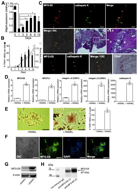
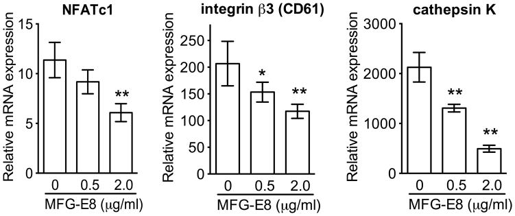
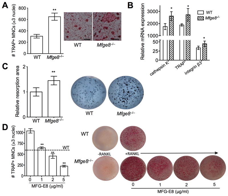
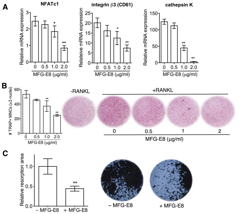
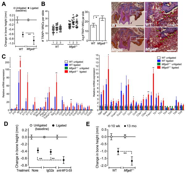
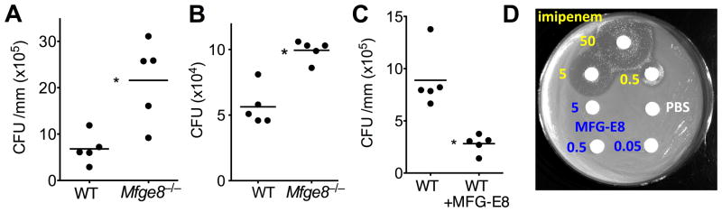
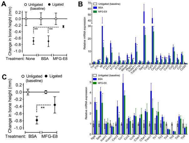
The glycoprotein milk fat globule-epidermal growth factor factor 8 (MFG-E8) is expressed in several tissues and mediates diverse homeostatic functions. However, whether it plays a role in bone homeostasis has not been established. In this study, we show for the first time, to our knowledge, that osteoclasts express and are regulated by MFG-E8. Bone marrow-derived osteoclast precursors from MFG-E8-deficient (Mfge8(-/-)) mice underwent increased receptor activator of NF-κB ligand-induced osteoclastogenesis, leading to enhanced resorption pit formation compared with wild-type controls. Consistently, exogenously added MFG-E8 inhibited receptor activator of NF-κB ligand-induced osteoclastogenesis from mouse or human osteoclast precursors. Upon induction of experimental periodontitis, an oral inflammatory disease characterized by loss of bone support of the dentition, Mfge8(-/-) mice exhibited higher numbers of osteoclasts and more bone loss than did wild-type controls. Accordingly, local microinjection of anti-MFG-E8 mAb exacerbated periodontal bone loss in wild-type mice. Conversely, microinjection of MFG-E8 inhibited bone loss in experimental mouse periodontitis. In comparison with wild-type controls, Mfge8(-/-) mice also experienced >60% more naturally occurring chronic periodontal bone loss. In conclusion, MFG-E8 is a novel homeostatic regulator of osteoclasts that could be exploited therapeutically to treat periodontitis and perhaps other immunological disorders associated with inflammatory bone loss.


Figures

Figure 1
Figure 1. Expression of MFG-E8 in the periodontal tissue and by in vitro generated OCLs
(A) Timecourse of MFG-E8 mRNA expression in the periodontal tissue after ligature-induced periodontitis. Results (means ± SD; n = 5 mice) were normalized to GAPDH mRNA and presented relative to those at d0, set as 1. (B) Mice were treated as above and TRAP+ MNCs were enumerated and averaged (with SD) from 60 random coronal sections (20 from each of three mice per group) of ligated teeth with surrounding periodontal tissue. Dashed line with open circles shows timecourse of bone loss in similarly treated mice (means ± SD; n = 5). (C) Tissue sections from ligature-induced periodontitis at d5 were stained as indicated. Bottom row images involve the same section processed for immunofluorescence and TRAP staining. B, bone; D, dentin; DIC, differential interference contrast; PL, periodontal ligament. Scale bars; 50μm (white), 500μm (black). (D) RANKL-induced OCLs from progenitor RAW264.7 cells (+RANKL) were assayed for mRNA expression of indicated molecules. Results (means ± SD; n = 3) were normalized to GAPDH mRNA and presented relative to those of undifferentiated RAW264.7 cells (−RANKL), set as 1. (E) Light microscopy of undifferentiated (−RANKL) and RANKL-differentiated RAW264.7 cells (scale bar, 50μm) and enumeration of TRAP+ MNCs in the cultures (means ± SD; n = 3). (F) DIC and fluorescent images of RANKL-differentiated OCL stained for MFG-E8 and nuclei (DAPI) (scale bar, 50μm). (G) Anti-MFG-E8 immunoblotting of cell lysates from undifferentiated RAW264.7 cells (−RANKL) and differentiated OCLs (+RANKL). (H) Immunoprecipitation of MFG-E8 from culture supernatants of RANKL-differentiated OCLs using goat anti-MFG-E8 IgG antibody (1, OCLs differentiated from RAW264.7 cells; 2, OCLs differentiated from mouse BM-derived precursors) followed by immunoblotting with the same antibody. *P < 0.01. NS, not significant.
Figure 2
Figure 2. MFG-E8 inhibits RANKL-induced differentiation of RAW264.7 OCLs
RANKL-induced osteoclastogenesis from progenitor RAW264.7 cells was performed in the absence or presence of the indicated concentrations of MFG-E8 and mRNA expression of the indicated molecules was assayed by qPCR. Results (means ± SD; n = 4) were normalized to GAPDH mRNA and presented relative to those of undifferentiated RAW264.7 cells, set as 1. *P < 0.05; **P < 0.01.
Figure 3
Figure 3. MFG-E8 regulates osteoclast differentiation and function
(A) Enumeration of TRAP+ MNCs in RANKL-stimulated cultures of WT and Mfge8−/− OCPs and representative photomicrographs (scale bar, 100μm). (B) RANKL-induced OCLs were generated from WT or Mfge8−/− OCPs and after 3d were assayed for mRNA expression of the indicated molecules. Results were normalized to those of GAPDH mRNA and presented relative to those of undifferentiated OCPs, set as 1. (C) WT and Mfge8−/− OCPs were cultured under osteoclastogenic conditions for 4d on Ca3(PO4)2-coated wells and the total resorption area (dark spots) in each culture was measured and expressed relative to the WT group, set as 1. (D) RANKL-induced osteoclastogenesis from Mfge8−/− OCPs in the presence of increasing MFG-E8 concentrations. TRAP+ MNCs were counted (left). Dashed line marks the number of OCLs formed from WT OCPs (no exogenous MFG-E8 added). (D) Data are means ± SD (A, B, and D, n = 3; C, n = 6). *P < 0.05, **P < 0.01 compared to WT or untreated control.
Figure 4
Figure 4. Regulation of human osteoclastogenesis and resorption pit formation by MFG-E8
Human CD14+ monocytes underwent RANKL-induced osteoclastogenesis, as detailed in Methods, in the presence of the indicated increasing concentrations of MFG-E8. (A) The mRNA expression of the indicated molecules was assayed by qPCR. Results were normalized to GAPDH mRNA and presented relative to those of undifferentiated monocytes (not treated with RANKL), assigned an average value of 1. (B) Cells were stained for TRAP expression and TRAP+ MNCs were counted (left). −RANKL indicates control monocytes not subjected to osteoclastogenesis. (C) CD14+ monocytes on Ca3(PO4)2-coated wells were cultured under osteoclastogenic conditions for 5d and resorptive areas (dark spots) were visualized by light microscopy. The total resorbed area in each culture was measured and expressed relative to the WT group, set as 1 (left). Data are means ± SD (A and B, n = 3; C, n = 8). *P < 0.05, **P < 0.01 compared to untreated control.
Figure 5
Figure 5. MFG-E8 regulates osteoclastogenesis and bone loss in vivo
(A) Periodontal bone loss was induced for 5d in WT or Mfge8−/− mice by ligating a maxillary second molar and leaving the contralateral tooth unligated (baseline control). (B) WT and Mfge8−/− mice were treated as above and TRAP+ MNCs were enumerated from 20 random coronal sections of the ligated molar from each of three mice (left) and averaged with SD from the total 60 sections per group (middle). Arrows in sections stained with TRAP, hematoxylin, orange-G, and aniline blue indicate OCLs adjacent to bone (right). Scale bars; 100μm (white), 500μm (black). (C) Dissected gingiva from mice used in A were processed for qPCR to determine mRNA expression of indicated molecules. Results were normalized to GAPDH mRNA and presented as fold change in the transcript levels in ligated sites relative to those of unligated sites (assigned an average value of 1). (D) Periodontal bone loss in mice locally microinjected with 5 μg anti-MFG-E8 mAb or IgG2a control 1d before placing the ligature and every day thereafter until the day before sacrifice (d5). (E) Naturally-occurring bone loss in 13-month-old Mfge8−/− mice and age-matched WT controls relative to bone measurements in 10-wk-old WT mice (0 baseline). Data are means ± SD (n = 5–8 mice per group, except for B; n = 3). *P < 0.05, **P < 0.01 compared with control or between indicated groups.
Figure 6
Figure 6. MFG-E8 reduces the periodontal bacterial burden without exerting direct antimicrobial activity
Periodontal microbiota counts were determined in WT or Mfge8−/− mice subjected to ligature-induced periodontitis (A) or to naturally-occurring periodontitis until the age of 13 months (B), as well as in WT mice in which ligature-induced periodontitis was performed with or without local treatment with 2.5 μg MFG-E8 (C). In A and C, bacteria were extracted from recovered ligatures and serial dilutions of bacterial suspensions were plated onto blood agar plates for anaerobic growth and CFU enumeration. In B, oral swabs held against the gumlines were taken and bacteria were cultured anaerobically for CFU enumeration as above. Each symbol represents an individual mouse and small horizontal lines indicate the mean. *P < 0.01. (D) Possible antimicrobial activity of MFG-E8 against mouse periodontal bacteria was determined by the disk inhibition zone method, using PBS and imipenem as negative and positive control, respectively. The experiment shown is representative of a total of 15 bacterial isolates and MFG-E8 consistently failed to inhibit bacterial growth. Numbers shown refer to μg of compound used.
Figure 7
Figure 7. rMFG-E8 inhibits ligature-induced periodontal bone loss in vivo
(A) Periodontal bone loss in WT mice locally microinjected with 2.5 μg MFG-E8 or BSA control 1d before placing the ligature and every day thereafter until the day before sacrifice (d5). (B) Dissected gingiva from mice used in A were processed to determine mRNA expression of the indicated molecules using qPCR. Results were normalized to GAPDH mRNA and presented as fold change in the transcript levels in ligated sites relative to those of unligated sites, set as 1. (C) Periodontal bone loss in Mfge8−/− mice locally microinjected with 2.5 μg MFG-E8 or BSA control as outlined in A. Data are means ± SD (n = 5–6 mice per group). *P < 0.05, **P < 0.01 compared with control or between indicated groups.

Similar articles

Cited by

References

    1. Raymond A, Ensslin MA, Shur BD. SED1/MFG-E8: a bi-motif protein that orchestrates diverse cellular interactions. J Cell Biochem. 2009;106:957–966. - PMC - PubMed
    1. Aziz M, Jacob A, Matsuda A, Wang P. Review: milk fat globule-EGF factor 8 expression, function and plausible signal transduction in resolving inflammation. Apoptosis. 2011;16:1077–1086. - PubMed
    1. Hanayama R, Tanaka M, Miwa K, Shinohara A, Iwamatsu A, Nagata S. Identification of a factor that links apoptotic cells to phagocytes. Nature. 2002;417:182–187. - PubMed
    1. Bu HF, Zuo XL, Wang X, Ensslin MA, Koti V, Hsueh W, Raymond AS, Shur BD, Tan XD. Milk fat globule-EGF factor 8/lactadherin plays a crucial role in maintenance and repair of murine intestinal epithelium. J Clin Invest. 2007;117:3673–3683. - PMC - PubMed
    1. Aziz MM, Ishihara S, Mishima Y, Oshima N, Moriyama I, Yuki T, Kadowaki Y, Rumi MA, Amano Y, Kinoshita Y. MFG-E8 attenuates intestinal inflammation in murine experimental colitis by modulating osteopontin-dependent alphavbeta3 integrin signaling. J Immunol. 2009;182:7222–7232. - PubMed

Publication types

MeSH terms