

Case Reports
doi: 10.4103/0973-029X.131950.
Branchial cleft cyst: A case report and review of literature
Affiliations
- PMID: 24959062
- PMCID: PMC4065440
- DOI: 10.4103/0973-029X.131950
Item in Clipboard
Case Reports
Branchial cleft cyst: A case report and review of literature
J Oral Maxillofac Pathol.
2014 Jan.
Abstract
First branchial cleft anomaly is a rare disease of the head and neck. Because of its rarity, first branchial cleft anomaly is often misdiagnosed and results in inappropriate management. In this article, we present a case of type II first branchial cleft anomaly. A middle-aged woman who had suffered from swelling on lower jaw visited our department with the chief complaint of a swelling. She underwent complete excision of the lesion with preservation of the facial nerve. The patient recovered well and had no recurrence at 1-year of follow up.
Keywords: Branchial cleft cyst; cervical lymphoepithelial cyst; first branchial anomaly.
Conflict of interest statement
Figures

Clinical image showing swelling in the neck

Gross specimen of the excised lesion

Photomicrograph showing cystic lesion lined by stratified squamous epithelium. (H&E stain, ×40)
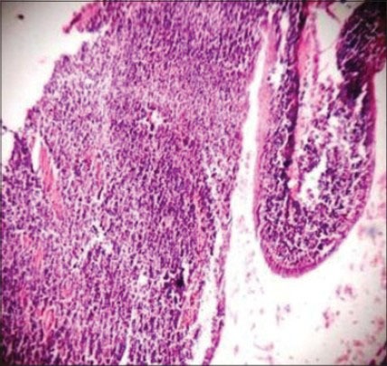
Photomicrograph showing cystic cavity lined by thin stratified squamous epithelium with the underlying connective tissue showing abundant lymphoid tissue aggregate (H&E stain, ×40)

Photomicrograph showing the germinal center within the lymphoid aggregate (H&E stain, ×100)

Photomicrograph showing cystic lumen lined by thin stratified squamous epithelium with flat rete ridges and underlying connective tissue showing abundant lymphoid tissue aggregate (H&E stain, ×100).
References
-
- Wei YK, Wang KJ, Jou YL, Chang YH, Chou CS. Type II first branchial cleft anomaly - A case report. Tzu Chi Med J. 2005:357–60.
-
- Arndal H, Bonding P. First branchial cleft anomaly. Clin Otolaryngol Allied Sci. 1996;21:203–7. - PubMed
-
- D’Souza AR, Uppal HS, De R, Zeitoun H. Updating concepts of first branchial cleft defects: A literature review. Int J Pediatr Otorhinolaryngol. 2002;62:103–9. - PubMed
-
- Frazer JE. The nomenclature of diseased states caused by certain vestigial structures in the neck. Br J Surg. 1923;11:131–6.
Publication types
LinkOut - more resources
Full Text Sources
Other Literature Sources
