

A new case of syringocystadenocarcinoma papilliferum: a rare pathology for a wide-ranging comprehension
- PMID: 24959179
- PMCID: PMC4052556
- DOI: 10.1155/2014/453874
A new case of syringocystadenocarcinoma papilliferum: a rare pathology for a wide-ranging comprehension
Abstract
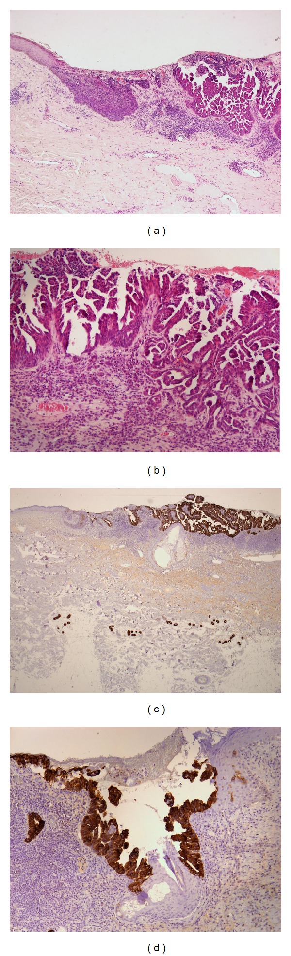
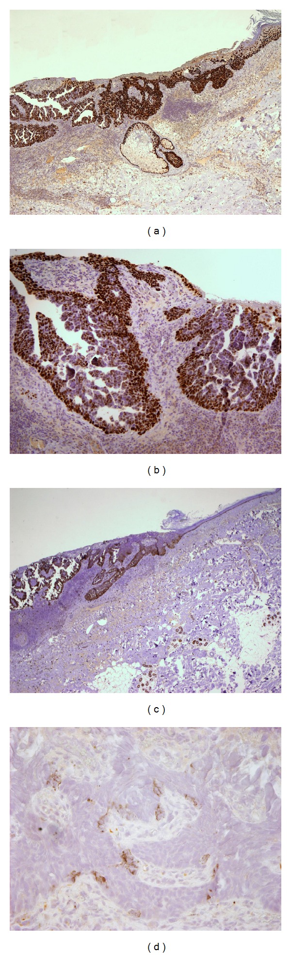
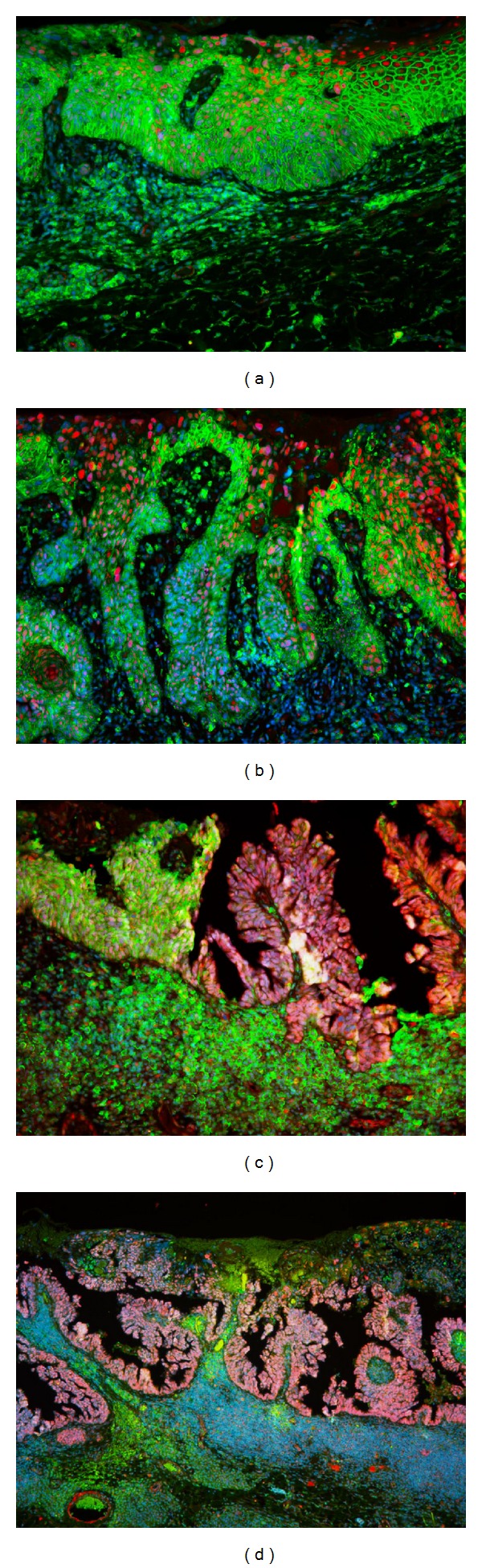
We report a new case of p63/cytokeratin 7 (CK7) positive syringocystadenocarcinoma papilliferum (SCACP), on the shoulder of an 88-year-old man, with superficial dermal infiltration and squamoid differentiation. We describe the 24th case of SCACP, the malignant counterpart of syringocystadenoma papilliferum (SCAP). At the present, we do not know whether SCACP arises from eccrine or apocrine glands because of the contrasting opinions in the literature. Only few histochemical and ultrastructural studies have previously advised that SCACP could arise from pluripotent stem cells. Through our case, we wish to suggest the stem cell-like properties of the syringocystadenocarcinoma papilliferum. This rare neoplasm shows two different patterns of stem cell marker expression in the glandular and squamous components, respectively. For the double phenotype of SCACP, we propose it like an intriguing model to study histogenesis and stem cell properties for more wide-ranging epithelial tumors.
Figures
References
-
- Hoekzema R, Leenarts MFE, Nijhuis EWP. Syringocystadenocarcinoma papilliferum in a linear nevus verrucosus. Journal of Cutaneous Pathology. 2011;38(2):246–250. - PubMed
-
- Leeborg N, Thompson M, Rossmiller S, Gross N, White C, Gatter K. Diagnostic pitfalls in syringocystadenocarcinoma papilliferum: case report and review of the literature. Archives of Pathology and Laboratory Medicine. 2010;134(8):1205–1209. - PubMed
-
- Yamamoto O, Doi Y, Hamada T, Hisaoka M, Sasaguri Y. An immunohistochemical and ultrastructural study of syringocystadenoma papilliferum. The British Journal of Dermatology. 2002;147(5):936–945. - PubMed
-
- Ishida-Yamamoto A, Sato K, Wada T, Takahashi H, Iizuka H. Syringocystadenocarcinoma papilliferum: case report and immunohistochemical comparison with its benign counterpart. Journal of the American Academy of Dermatology. 2001;45(5):755–759. - PubMed
-
- Dissanayake RV, Salm R. Sweat-gland carcinomas: prognosis related to histological type. Histopathology. 1980;4(4):445–466. - PubMed
LinkOut - more resources
Full Text Sources
Other Literature Sources
Research Materials
