. 2014 Jul;92(7):598-604.
doi: 10.1139/cjpp-2013-0490. Epub 2014 Jan 31.

Hyperglycemia enhances function and differentiation of adult rat cardiac fibroblasts

Affiliations

Hyperglycemia enhances function and differentiation of adult rat cardiac fibroblasts

Patricia E Shamhart et al. Can J Physiol Pharmacol. 2014 Jul.

Abstract

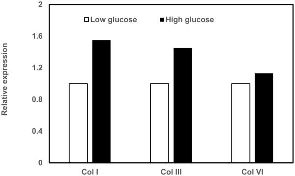
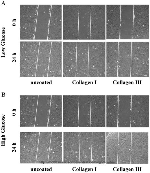
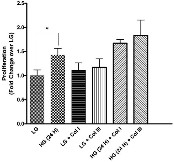
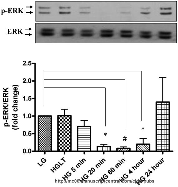
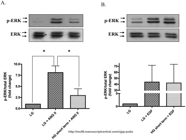
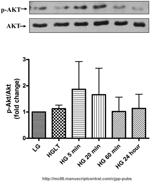
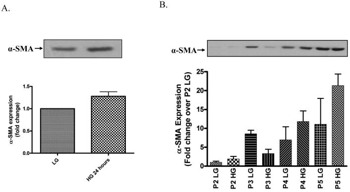
Diabetes is an independent risk factor for cardiovascular disease that can eventually cause cardiomyopathy and heart failure. Cardiac fibroblasts (CF) are the critical mediators of physiological and pathological cardiac remodeling; however, the effects of hyperglycemia on cardiac fibroblast function and differentiation is not well known. Here, we performed a comprehensive investigation on the effects of hyperglycemia on cardiac fibroblasts and show that hyperglycemia enhances cardiac fibroblast function and differentiation. We found that high glucose treatment increased collagen I, III, and VI gene expression in rat adult cardiac fibroblasts. Interestingly, hyperglycemia increased CF migration and proliferation that is augmented by collagen I and III. Surprisingly, we found that short term hyperglycemia transiently inhibited ERK1/2 activation but increased AKT phosphorylation. Finally, high glucose treatment increased spontaneous differentiation of cardiac fibroblasts to myofibroblasts with increasing passage compared with low glucose. Taken together, these findings suggest that hyperglycemia induces cardiac fibrosis by modulating collagen expression, migration, proliferation, and differentiation of cardiac fibroblasts.

Keywords: cardiac fibroblast; collagen; collagène; concentration élevée de glucose; diabetes; diabète; fibroblaste cardiaque; high glucose; migration; myofibroblast; myofibroblastes; proliferation; prolifération.


Figures

Figure 1
Figure 1. RT-PCR analysis of collagens
Cardiac fibroblasts were cultured in either low glucose (5 mM) or high glucose (25 mM) and total RNA was isolated and subjected cDNA was synthesized using reverse transcription. The expression of collagen I, III, VI and GAPDH transcripts were measured in real-time PCR, normalized for GAPDH and expressed relative to low glucose conditions.
Figure 2
Figure 2. Hyperglycemia and Collagen types I and III potently promote cardiac fibroblast migration
Cardiac fibroblasts were plated onto collagen types I and III and tissue culture plates. Cells were grown to 90-100% confluence, serum-starved for 24 hours, and scratched with 200 μL pipet tip. The wounds were washed with PBS and fresh media with the appropriate glucose concentration was added to the cells. Panel A depicts increased fibroblast migration at 24 hours on both collagen types I and III in low glucose. Panel B reveals the compounded effects of hyperglycemia (high glucose) and collagen types I and III on cardiac fibroblast migration. Data are representative of triplicate wells from 4 separate CF preparations (n=4).
Figure 3
Figure 3. Hyperglycemia stimulates cardiac fibroblast proliferation
Cardiac fibroblasts were cultured in 96 well culture plates uncoated or coated with collagen I and III in the presence of low glucose (LG) and high glucose (HG) for 24 h. High glucose significantly induced fibroblast proliferation. Data are representative of 5 separate wells from 3 separate CF preparations (n=5). Statistical significance (p<0.05) between groups was determined by one-way ANOVA.
Figure 4
Figure 4. High glucose transiently inhibits basal ERK 1/2 phosphorylation
Cardiac fibroblasts were cultured in either low glucose (5 mM) or high glucose (25 mM) for the designated time and cell lysates were collected and subjected to Western blot analysis for phospho-ERK and total ERK. A) Basal ERK phosphorylation was lower in fibroblasts treated with high glucose for 20 min (0.13±0.06 fold, p<0.05), 60 min (0.08±0.42 fold, p<0.001), and 4 h (0.20±0.16 fold, p<0.05). Data are representative of duplicate wells from 4 separate CF preparations (n=4). Statistical significance (p<0.05) between groups was determined by one-way ANOVA.
Figure 5
Figure 5. High glucose inhibits ANG II-induced ERK 1/2 activation
CF were either culture in low glucose or high glucose and then stimulated with ANG II or EGF for 5 minutes. Cell lysates were collected and subjected to Western blot analysis for phospho-ERK and total ERK. A) Representative Western blot and quantitative analysis depicting that high glucose pretreatment attenuates ANG II-induced ERK1/2 phosphorylation (p<0.05). B) In contrast, high glucose does not prevent EGF-induced ERK 1/2 activation. Data are representative of duplicate from 4 separate CF preparations (n=4).
Figure 6
Figure 6. Short-term hyperglycemia increases AKT phsphorylation
CF were cultured in the indicated glucose concentrations for the time frame specified. The representative Western blot and quantitative analysis reveal that incubation with high glucose for 5 and 20 min increases AKT phosphorylation. Data are representative of duplicate wells from 4 separate CF preparations (n=4).
Figure 7
Figure 7. Hyperglycemia accelerates CFs differentiation to myofibroblasts
Cardiac fibroblasts were cultured in either low glucose (5 mM) or high glucose (25 mM) for the designated time frame and cell lysates were collected and subjected to Western blot analysis for a-SMA expression. Representative Western blot and summary graph for α-SMA expression following a 24 h high glucose incubation (A) and at each cell culture passage (B). Each Western blot was normalized to bands from the napthol blue staining of the same membrane. Each blot and summary graph are mean fold change ± SEM and representative duplicate wells from 2 separate CF preparations (n=2).

References

    1. Aragno M, Mastrocola R, Alloatti G, Vercellinatto I, Bardini P, Geuna S, Catalano MG, Danni O, Boccuzzi G. Oxidative stress triggers cardiac fibrosis in the heart of diabetic rats. Endocrinology. 2008;149:380–388. - PubMed
    1. Asbun J, Manso AM, Villarreal FJ. Profibrotic influence of high glucose concentration on cardiac fibroblast functions: effects of losartan and vitamin E. Am J Physiol Heart Circ Physiol. 2005;288:H227–H234. - PubMed
    1. Asbun J, Villarreal FJ. The Pathogenesis of Myocardial Fibrosis in the Setting of Diabetic Cardiomyopathy. Journal of American College of Cardiology. 2006;47:693–700. - PubMed
    1. Deckert T, Poulsen JE, Larsen M. Prognosis of diabetics with diabetes onset before the age of thirty-one. I. Survival, causes of death, and complications. Diabetologia. 1978;14:363–370. - PubMed
    1. Dorman JS, Laporte RE, Kuller LH, Cruickshanks KJ, Orchard TJ, Wagener DK, Becker DJ, Cavender DE, Drash AL. The Pittsburgh insulin-dependent diabetes mellitus (IDDM) morbidity and mortality study. Mortality results. Diabetes. 1984;33:271–276. - PubMed

Publication types