. 2014 Aug;124(8):3419-30.
doi: 10.1172/JCI74523. Epub 2014 Jun 24.

RNA-binding protein RBM20 represses splicing to orchestrate cardiac pre-mRNA processing

RNA-binding protein RBM20 represses splicing to orchestrate cardiac pre-mRNA processing

Henrike Maatz et al. J Clin Invest. 2014 Aug.

Abstract

Mutations in the gene encoding the RNA-binding protein RBM20 have been implicated in dilated cardiomyopathy (DCM), a major cause of chronic heart failure, presumably through altering cardiac RNA splicing. Here, we combined transcriptome-wide crosslinking immunoprecipitation (CLIP-seq), RNA-seq, and quantitative proteomics in cell culture and rat and human hearts to examine how RBM20 regulates alternative splicing in the heart. Our analyses revealed the presence of a distinct RBM20 RNA-recognition element that is predominantly found within intronic binding sites and linked to repression of exon splicing with RBM20 binding near 3' and 5' splice sites. Proteomic analysis determined that RBM20 interacts with both U1 and U2 small nuclear ribonucleic particles (snRNPs) and suggested that RBM20-dependent splicing repression occurs through spliceosome stalling at complex A. Direct RBM20 targets included several genes previously shown to be involved in DCM as well as genes not typically associated with this disease. In failing human hearts, reduced expression of RBM20 affected alternative splicing of several direct targets, indicating that differences in RBM20 expression may affect cardiac function. Together, these findings identify RBM20-regulated targets and provide insight into the pathogenesis of human heart failure.


Figures
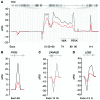
Figure 7
Figure 7. Splicing patterns of RBM20-regulated exons in individuals with differential endogenous RBM20 expression levels.
ΔPSI values for RBM20 regulated exons of (A) TTN, (B) RYR2, (C) CAMK2D, and (D) LDB3 are shown in red for low compared with high RBM20-expressing individuals. ΔPSI values for S635A compared with control subjects shown in gray serve as indicators of RBM20 regulated events.
Figure 6
Figure 6. Alignment of orthologous rat and human exons for Ryr2, Lmo7, Rtn4, and Pdlim3 to compare RBM20-regulated exon usage.
PSI scores for the RBM20-deficient individual (S635A) are indicated in red and the average of 5 control subjects from the human heart failure cohort (CP) in blue. The average PSI of 3 rats per genotype is shown below (WT in blue and Rbm20–/– in red). Resulting ΔPSIs are shown in black (S635A compared with CP) and in gray (WT compared with Rbm20–/– rats). Deflections in ΔPSI values in differentially spliced regions are highly conserved across species. (A) Ryr2 shows RBM20-dependent splicing of a 24-bp exon included in RBM20-deficient hearts. The magnified view shows RBM20 cluster flanking the differentially spliced rat Ryr2 exon. (B) In Lmo7, RBM20 deficiency causes retention of exons 9 and 10. The zoom-in shows RBM20 cluster flanking exon 10 immediately upstream of its 3′ splice site. (C) Rtn4 exons 3 and 4 are differentially spliced. The magnification shows the intronic RBM20 cluster flanking rat exons 3 and 4. (D) Pdlim3 exons 4–6 are mutually exclusive and differentially regulated by RBM20. The zoom-in shows the locations of RBM20 cluster upstream of exon 4.
Figure 5
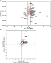
Figure 5. Impact of RBM20 S635A mutation on protein-protein interaction.
(A) WT versus S635A comparison. RBM20 was identified as highly and equally abundant in WT- and S635A-RBM20 samples, indicating successful immunoprecipitation. Red circles indicate spliceosomal interaction partners not affected by the mutation. Labeled red dots indicate spliceosomal interaction partners specific to WT- (log2 ratio > 0.4) or S635A-RBM20 (log2 ratio < –0.4). (B) Comparison of RNase-digested versus nondigested q-AP-MS identifies RNase-insensitive RBM20 interactors. Upper right quadrant contains WT- and lower left quadrant mutant-specific interactors enriched in RNase-digested and nondigested samples. Spliceosomal interaction partners are marked by red dots and labeled when WT- or mutant-specific in both protein-protein interaction screens. Bold labeled proteins were identified as also interacting with RBM20 in cardiomyocytes.
Figure 4
Figure 4. Identification of RBM20 protein-protein interaction partners.
(A) q-AP-MS using SILAC. Differentially SILAC-labeled HEK293 cells (light, medium, and heavy) were transfected with FLAG-tagged control vector, FLAG-tagged RBM20 WT, or S635A mutant. RBM20 protein complexes were enriched by affinity purification and analyzed by quantitative mass spectrometry. (B) The log2 ratios of protein-protein interactions for WT-RBM20 versus control are shown on the left. Red dots indicate known splicing factors. Validation of q-AP-MS hits by Western blotting is shown on the right. (C) U1–, U2–, and U4/U6.U5–specific proteins are grouped according to snRNP association and highlighted in blue when identified as interacting with RBM20.
Figure 3
Figure 3. RBM20 binds to intronic splicing silencers upstream and downstream of repressed exons.
(AC) ΔPSIs are shown as black lines (WT compared with homozygous rats). Elevated versus decreased ΔPSIs show inclusion or exclusion of exons, respectively. Red ticks indicate mapped RBM20 cluster positions. (A) Ttn is predominantly spliced in the elastic region. Mapped RBM20-binding sites coincide with differential splicing. (B) Camk2d exons 14–16 are mutually exclusive and regulated by RBM20. Mapped RBM20-binding sites are located upstream of exon 14 and downstream of exon 15. (C) Ldb3 undergoes an exon switch affecting exon 4 versus exons 5–7. Mapped binding sites are upstream of exon 5 and downstream of exon 8. (D) Mean CLIP density near RBM20 activated (red) and repressed exons (blue). Dotted line indicates density around exons not regulated by RBM20. Gray shows 90% CIs. (E) Right panel: specific activity of RBM20 on Ttn RNA from the PEVK region in a splice-reporter assay. Rbm20 expression leads to exclusion of the firefly luciferase–containing (Fluc) exon. Ratio of Fluc to Renilla luciferase (Rluc) activity (downstream exon) reflects splice activity. Reporter activity depends on mutation of 2 UCUU elements in cluster CID016226, but not outside the cluster. Mutations are indicated as X. White boxes only show native reporter. Controls: PEVK-construct cotransfected with empty vector (–), Rbm20 cotransfected with a Ttn M-band region construct not affected by RBM20 (M). ***P < 0.001 compared with native reporter. Left: EMSA evaluating RBM20 binding to Ttn PEVK-derived RNAs. White arrows indicate input RNA; black arrows indicate RNA-protein complex.
Figure 2
Figure 2. HITS-CLIP in rat cardiomyocytes.
(A) Transcriptome-wide distribution of HITS-CLIP–derived RBM20 target sites in cardiomyocytes. The ratio of HITS-CLIP to cardiac RNA-seq percentage coverage in each region is displayed. (B) Binding motif enrichment in cardiomyocyte Rbm20 clusters. The log10 frequencies of 5-mers in clusters correlated with frequencies of 5-mers in control sequence. (C) Sequence logo for the RBM20 RRE in cardiomyocytes was computed from the top 1000 intronic binding sites using MEME. (D) Confirmation of alternative splicing of genes with high ΔPSI values as determined by RNA-seq. i1 indicates isoforms expressed at higher levels in WT than in mutant rats and i2 those expressed at higher levels in mutant than in WT rats. Rbm20 was amplified as control and verifies the indicated Rbm20 genotype of the samples tested. (E) ΔPSI values > |0.01| of RBM20-regulated exons in WT versus Rbm20-deficient rat hearts. RBM20 acts predominantly as a splicing repressor in cardiomyocytes. (F) RBM20-regulated versus expected cardiac splicing events. MXE, mutually exclusive exon; CE, cassette exon; CNE, constitutive exon.
Figure 1
Figure 1. PAR-CLIP in HEK293 cells identifies RBM20 RRE.
(A) Transcriptome-wide distribution of RBM20 PAR-CLIP consensus library clusters. The ratio of PAR-CLIP to control HEK293 RNA-seq percentage coverage in each region is displayed. (B) Binding motif enrichment in RBM20 PAR-CLIP libraries. The log2 enrichments of 5-mers in cluster-centered regions of the 4SU and 6SG libraries are correlated. UCUU core–containing 5-mers were most abundant. (C) Sequence logo for the RBM20 RRE was computed from the top 1000 intronic binding sites of the consensus library using MEME. (D) Positional transition frequency for intronic PAR-CLIP clusters anchored at the UCUU core of the RBM20 RRE (black line) and control CCRs randomly placed in the same intron (blue line). (E) Phosphorimage of native PAGE resolving complexes of immunoprecipitated full-length RBM20 protein with WT and mutated Ryr2 target RNA oligonucleotides.

References

    1. Chen M, Manley JL. Mechanisms of alternative splicing regulation: insights from molecular and genomics approaches. Nat Rev Mol Cell Biol. 2009;10(11):741–754. - PMC - PubMed
    1. Cartegni L, Chew SL, Krainer AR. Listening to silence and understanding nonsense: exonic mutations that affect splicing. Nat Rev Genet. 2002;3(4):285–298. doi: 10.1038/nrg775. - DOI - PubMed
    1. Caceres JF, Kornblihtt AR. Alternative splicing: multiple control mechanisms and involvement in human disease. Trends Genet. 2002;18(4):186–193. doi: 10.1016/S0168-9525(01)02626-9. - DOI - PubMed
    1. Douglas AG, Wood MJ. RNA splicing: disease and therapy. Brief Funct Genomics. 2011;10(3):151–164. doi: 10.1093/bfgp/elr020. - DOI - PubMed
    1. Guo W, et al. RBM20, a gene for hereditary cardiomyopathy, regulates titin splicing. Nat Med. 2012;18(5):766–773. doi: 10.1038/nm.2693. - DOI - PMC - PubMed

Publication types

MeSH terms

Substances