. 2014 Aug;124(8):3529-39.
doi: 10.1172/JCI74994. Epub 2014 Jun 24.

KLHL40 deficiency destabilizes thin filament proteins and promotes nemaline myopathy

KLHL40 deficiency destabilizes thin filament proteins and promotes nemaline myopathy

Ankit Garg et al. J Clin Invest. 2014 Aug.

Abstract

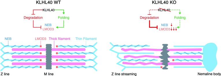
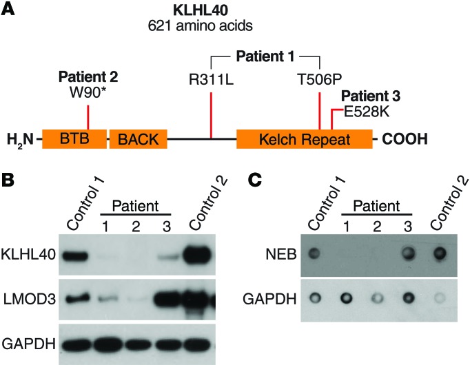
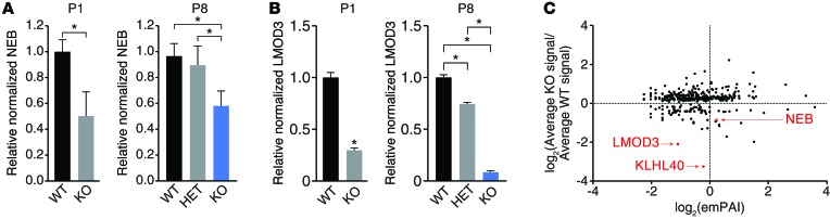
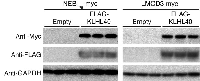
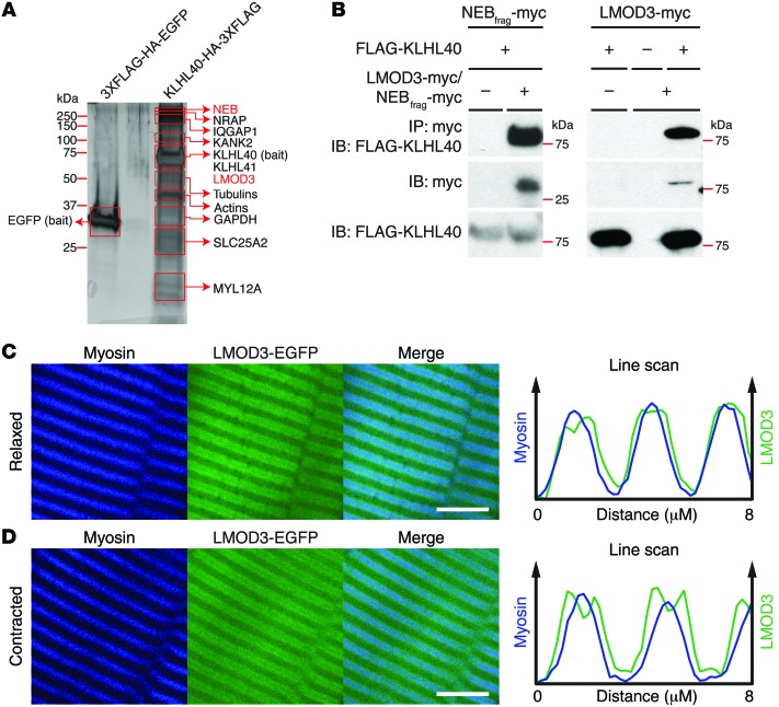
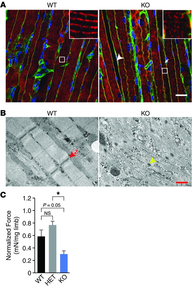
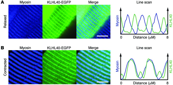
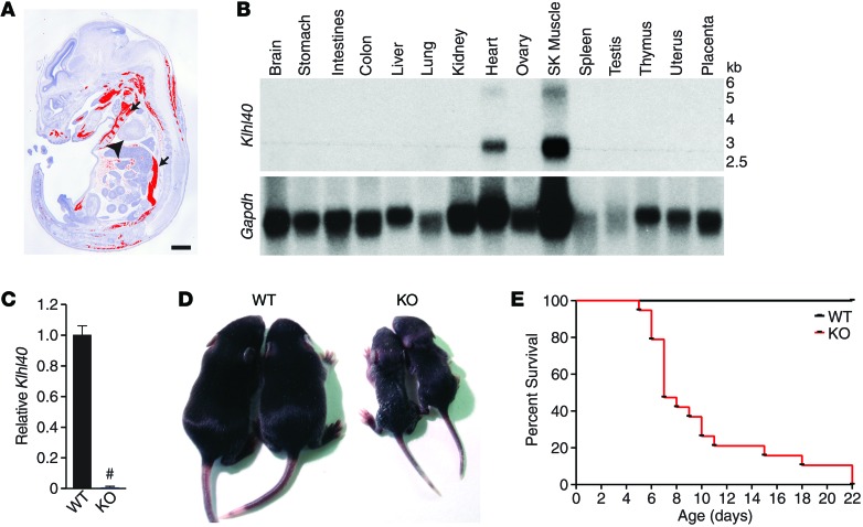
Nemaline myopathy (NM) is a congenital myopathy that can result in lethal muscle dysfunction and is thought to be a disease of the sarcomere thin filament. Recently, several proteins of unknown function have been implicated in NM, but the mechanistic basis of their contribution to disease remains unresolved. Here, we demonstrated that loss of a muscle-specific protein, kelch-like family member 40 (KLHL40), results in a nemaline-like myopathy in mice that closely phenocopies muscle abnormalities observed in KLHL40-deficient patients. We determined that KLHL40 localizes to the sarcomere I band and A band and binds to nebulin (NEB), a protein frequently implicated in NM, as well as a putative thin filament protein, leiomodin 3 (LMOD3). KLHL40 belongs to the BTB-BACK-kelch (BBK) family of proteins, some of which have been shown to promote degradation of their substrates. In contrast, we found that KLHL40 promotes stability of NEB and LMOD3 and blocks LMOD3 ubiquitination. Accordingly, NEB and LMOD3 were reduced in skeletal muscle of both Klhl40-/- mice and KLHL40-deficient patients. Loss of sarcomere thin filament proteins is a frequent cause of NM; therefore, our data that KLHL40 stabilizes NEB and LMOD3 provide a potential basis for the development of NM in KLHL40-deficient patients.


Figures

Figure 8
Figure 8. Proposed mechanism of NM pathogenesis due to loss of KLHL40.
Under normal conditions, KLHL40 either blocks the degradation or promotes successful folding of NEB and LMOD3, which function to stabilize the thin filament, allowing normal sarcomere function. In muscles lacking KLHL40, NEB and LMOD3 protein levels are reduced, resulting in destabilization of thin filaments, sarcomere dysfunction, and subsequent NM.
Figure 7
Figure 7. Analyzing LMOD3 and NEB expression in KLHL40-deficient patients.
(A) Relative location of KLHL40 mutations in patients analyzed in B and C. Note that patient 1 is a compound heterozygote for the indicated mutations. (B) Detection of LMOD3 and KLHL40 by Western blot analysis and (C) NEB by dot blot analysis in control and KLHL40-deficient patient skeletal muscle biopsies. Patients 1 and 2 have a clear decrease in LMOD3 and NEB compared with age-matched control, control 1. Patient 3 compared with age-matched control, control 2, does not have decreased LMOD3, and changes in NEB are inconclusive due to issues with GAPDH signal. Control 1 and control 2 are 11-day-old and 6-month-old neonate controls, respectively. Patient 1 was biopsied at 2 days, patient 2 was biopsied at 25 days, and patient 3 was biopsied at 3 months of age. GAPDH is shown for loading control.
Figure 6
Figure 6. Reduced NEB and LMOD3 in KLHL40-deficient muscles.
(A and B) Densitometry analysis of P1 and P8 quadriceps muscle (A) NEB dot blot and (B) LMOD3 Western blot shows downregulation of both proteins in KO muscles compared with WT muscles (see Supplemental Figure 14) (P1: WT and KO, n = 3; P8: WT, n = 5, HET, n = 4, and KO, n = 5). *P < 0.05, FDR = 0.05. Data are presented as mean ± SEM. (C) Quantitative proteomic analysis of relative protein changes between P6 WT (n = 3) and KO (n = 3) whole skeletal muscle shows that LMOD3 and NEB are 2 of the most downregulated proteins without corresponding changes in transcription (Supplemental Figure 14G). Data are arbitrarily stratified by exponentially modified protein abundance index (emPAI) to allow for better resolution of individual data points. Only proteins with significant (P < 0.05) changes between WT and KO mice are shown. Values for each point are listed in Supplemental Table 2.
Figure 5
Figure 5. KLHL40 increases NEBfrag and LMOD3 protein levels.
Western blot analysis of COS7 cells expressing NEBfrag-myc or LMOD3-myc without (Empty) or with FLAG-KLHL40. GAPDH is used as a loading control.
Figure 4
Figure 4. KLHL40 binds 2 thin filament proteins, NEB and LMOD3, and LMOD3 localizes to the sarcomere A band.
(A) Top KLHL40 binding partners identified following TAP of KLHL40 from cultured C2C12 myotubes. A representative silver-stained gel with TAP protein from myotubes infected with 3XFLAG-HA-EGFP (negative control) or KLHL40-HA-3XFLAG is shown. Proteins listed next to each box indicate the most abundant protein(s) of the correct molecular weight identified in each corresponding area. Abundance is based on estimated enrichment using exponentially modified protein abundance index. TAP of 3XFLAG-HA-KLHL40 protein was also analyzed, and the same top-binding partners were identified (Supplemental Figure 9). Proteins shown in red (NEB and LMOD3) indicate KLHL40 binding partners mutually identified in the TAP and yeast 2-hybrid experiment (Table 1). (B) Coimmunoprecipitation of FLAG-KLHL40 with LMOD3-myc and NEBfrag-myc from COS7 cells. “–” refers to empty FLAG vector for FLAG-KLHL40 and empty myc vector for either LMOD3-myc or NEBfrag-myc. (C and D) LMOD3-EGFP localization relative to myosin in (C) relaxed muscles and (D) contracted muscles shows that LMOD3 resides in the sarcomere A band. Graphs are representative line scans of indicated signals calculated over 8 μm of distance along the length of the myofiber. Scale bar: 5 μm.
Figure 3
Figure 3. KO mice have disrupted muscle structure and function.
(A) Longitudinal sections of P8 diaphragm muscles from WT and KO mice stained with desmin (red), DAPI (blue), and wheat germ agglutinin (green). The white arrow indicates disrupted myofibers. The white arrowhead denotes fibers staining abnormally for desmin that are more frequent in, but not specific to, KO muscles. Insets show normal striated pattern of Z lines in WT muscles but that some KO fibers have complete loss of sarcomere organization. Scale bar: 20 μm. (B) Electron microscopy analysis of longitudinal sections of P8 diaphragms from WT and KO mice shows some highly disorganized fibers. The red “Z” with red arrowhead indicates representative Z line in WT section. The yellow arrowhead denotes representative nemaline-like body. Scale bar: 1 μm. (C) Maximum contractile force of P1 WT, Klhl40+/– (HET), and KO hind limb following 150-Hz stimulation, normalized to limb mass. KO muscle shows greater than 50% loss of force compared with that of both WT and HET mice (WT, n = 6; HET, n = 17; and KO, n = 8). *P < 0.05, FDR = 0.05. Data are presented as mean ± SEM.
Figure 2
Figure 2. KLHL40 localizes to sarcomere I band and A band.
KLHL40-EGFP localization relative to myosin in (A) relaxed muscles and (B) contracted muscles shows that KLHL40 localizes to the I band and A band. Graphs are representative line scans of indicated signals calculated over 8 μm of distance along the length of the myofiber. Scale bar: 5 μm.
Figure 1
Figure 1. Klhl40 is expressed specifically in skeletal muscle and heart, and deletion of Klhl40 causes neonatal lethality.
(A) Sagittal sections of E15 mouse embryos were probed for Klhl40 mRNA using antisense radioisotopic probes. Signal for Klhl40 (pseudocolored red) only appears in developing muscle. Black arrows point to representative developing intercostal and back muscles from rostral to caudal, respectively. The black arrowhead denotes heart. Scale bar: 1 mm. (B) Northern blot of adult mouse tissues for Klhl40 and Gapdh (loading control) shows Klhl40 expression in skeletal muscle and heart. Numbers on right indicate size of transcripts in kilobases. SK Muscle, skeletal muscle. (C) qPCR analysis of Klhl40 transcript in P8 quadriceps in mice of the indicated genotypes. Values are normalized to 18S ribosomal RNA (n = 3 for both genotypes). #P < 0.05. (D) Representative image of surviving P8 KO pups with WT littermates. (E) Survival curve of WT versus KO mice shows early neonatal lethality in KO mice (WT, n = 21, and KO, n = 17).

References

    1. Wallgren-Pettersson C, Sewry CA, Nowak KJ, Laing NG. Nemaline myopathies. Semin Pediatr Neurol. 2011;18(4):230–238. doi: 10.1016/j.spen.2011.10.004. - DOI - PubMed
    1. Shimomura C, Nonaka I. Nemaline myopathy: comparative muscle histochemistry in the severe neonatal, moderate congenital, and adult-onset forms. Pediatr Neurol. 1989;5(1):25–31. doi: 10.1016/0887-8994(89)90005-2. - DOI - PubMed
    1. Laing NG, et al. A mutation in the α tropomyosin gene TPM3 associated with autosomal dominant nemaline myopathy NEM1. Nat Genet. 1995;10(2):249. - PubMed
    1. Lehtokari VL, et al. Identification of 45 novel mutations in the nebulin gene associated with autosomal recessive nemaline myopathy. Hum Mutat. 2006;27(9):946–956. doi: 10.1002/humu.20370. - DOI - PubMed
    1. Nowak KJ, et al. Mutations in the skeletal muscle α-actin gene in patients with actin myopathy and nemaline myopathy. Nat Genet. 1999;23(2):208–212. doi: 10.1038/13837. - DOI - PubMed

Publication types

MeSH terms