

Keratinocyte detachment-differentiation connection revisited, or anoikis-pityriasi nexus redux
- PMID: 24960166
- PMCID: PMC4069014
- DOI: 10.1371/journal.pone.0100279
Keratinocyte detachment-differentiation connection revisited, or anoikis-pityriasi nexus redux
Abstract
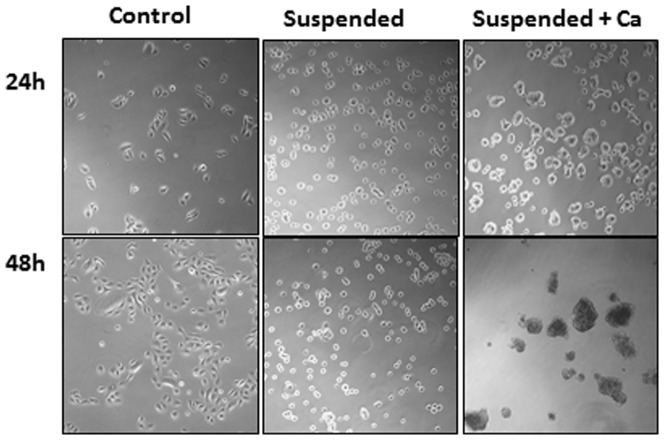
Epidermis, a continuously self-renewing and differentiating organ, produces a protective stratum corneum that shields us from external chemical, physical and microbial threats. Epidermal differentiation is a multi-step process regulated by influences, some unknown, others insufficiently explored. Detachment of keratinocytes from the basement membrane is one such pro-differentiation stimulus. Here, we define the transcriptional changes during differentiation, especially those caused by detachment from the substratum. Using comprehensive transcriptional profiling, we revisited the effects of detachment as a differentiation signal to keratinocytes. We identified the genes regulated by detachment, the corresponding ontological categories and, using metaanalysis, compared the genes and categories to those regulated by other pro-differentiating stimuli. We identified 762 genes overexpressed in suspended keratinocyte, including known and novel differentiation markers, and 1427 in attached cells, including basal layer markers. Detachment induced epidermis development, cornification and desmosomal genes, but also innate immunity, proliferation inhibitors, transcription regulators and MAPKs; conversely the attached cells overexpressed cell cycle, anchoring, motility, splicing and mitochondrial genes, and both positive and negative regulators of apoptosis. Metaanalysis identified which detachment-regulated categories overlap with those induced by suprabasal location in vivo, by reaching confluency in vitro, and by inhibition of JUN kinases. Attached and in vivo basal cells shared overexpression of mitochondrial components. Interestingly, melanosome trafficking components were also overexpressed in the attached and in vivo basal keratinocytes. These results suggest that specific pro-differentiation signals induce specific features of the keratinization process, which are in vivo orchestrated into harmonious epidermal homeostasis.
Conflict of interest statement
Figures
Similar articles
-
Integration of transcriptomics and spatial biology analyses reveals Galactomyces ferment filtrate promotes epidermal interconnectivity via induction of keratinocyte differentiation, proliferation and cellular bioenergetics.Int J Cosmet Sci. 2024 Dec;46(6):927-940. doi: 10.1111/ics.12991. Epub 2024 Jun 24. Int J Cosmet Sci. 2024. PMID: 38924095
-
ATP2C1 is specifically localized in the basal layer of normal epidermis and its depletion triggers keratinocyte differentiation.J Dermatol Sci. 2006 Jul;43(1):21-33. doi: 10.1016/j.jdermsci.2006.03.003. Epub 2006 Apr 18. J Dermatol Sci. 2006. PMID: 16621454
-
Selective changes in laminin adhesion and alpha 6 beta 4 integrin regulation are associated with the initial steps in keratinocyte maturation.Cell Growth Differ. 1996 May;7(5):615-28. Cell Growth Differ. 1996. PMID: 8732671
-
The epidermal keratinocyte as a model for the study of gene regulation and cell differentiation.Physiol Rev. 1997 Apr;77(2):397-424. doi: 10.1152/physrev.1997.77.2.397. Physiol Rev. 1997. PMID: 9114819 Review.
-
Polycomb group proteins are key regulators of keratinocyte function.J Invest Dermatol. 2011 Feb;131(2):295-301. doi: 10.1038/jid.2010.318. Epub 2010 Nov 18. J Invest Dermatol. 2011. PMID: 21085188 Free PMC article. Review.
Cited by
-
Regulation of integrin and extracellular matrix genes by HNRNPL is necessary for epidermal renewal.PLoS Biol. 2021 Sep 20;19(9):e3001378. doi: 10.1371/journal.pbio.3001378. eCollection 2021 Sep. PLoS Biol. 2021. PMID: 34543262 Free PMC article.
-
Dual Role of Act1 in Keratinocyte Differentiation and Host Defense: TRAF3IP2 Silencing Alters Keratinocyte Differentiation and Inhibits IL-17 Responses.J Invest Dermatol. 2017 Jul;137(7):1501-1511. doi: 10.1016/j.jid.2016.12.032. Epub 2017 Mar 6. J Invest Dermatol. 2017. PMID: 28274739 Free PMC article.
-
A novel ATAC-seq approach reveals lineage-specific reinforcement of the open chromatin landscape via cooperation between BAF and p63.Genome Biol. 2015 Dec 18;16:284. doi: 10.1186/s13059-015-0840-9. Genome Biol. 2015. PMID: 26683334 Free PMC article.
-
T helper 2 cell-directed immunotherapy eliminates precancerous skin lesions.J Clin Invest. 2025 Jan 2;135(1):e183274. doi: 10.1172/JCI183274. J Clin Invest. 2025. PMID: 39744942 Free PMC article.
-
Proteomics Characterization of Primary Human Oral Epithelial Cells Using a Novel Culture Technique for Use in Tissue Regeneration.MOJ Proteom Bioinform. 2015;2(4):52. doi: 10.15406/mojpb.2015.02.00052. Epub 2015 Aug 12. MOJ Proteom Bioinform. 2015. PMID: 27042699 Free PMC article.
References
-
- Lopez-Pajares V, Yan K, Zarnegar BJ, Jameson KL, Khavari PA (2013) Genetic pathways in disorders of epidermal differentiation. Trends Genet 29: 31–40 doi:10.1016/j.tig.2012.1010.1005 - DOI - PMC - PubMed
-
- Feingold KR, Elias PM (2013) Role of lipids in the formation and maintenance of the cutaneous permeability barrier. Biochim Biophys Acta 18: 00249–00247. - PubMed
-
- Perdigoto CN, Valdes VJ, Bardot ES, Ezhkova E (2014) Epigenetic regulation of epidermal differentiation. Cold Spring Harb Perspect Med 4(2): a015263 doi:015210.011101/cshperspect.a015263 - PMC - PubMed
-
- Bertrand-Vallery V, Belot N, Dieu M, Delaive E, Ninane N, et al. (2010) Proteomic profiling of human keratinocytes undergoing UVB-induced alternative differentiation reveals TRIpartite Motif Protein 29 as a survival factor. PLoS One 5: e10462 doi:10410.11371/journal.pone.0010462 - PMC - PubMed
Publication types
MeSH terms
LinkOut - more resources
Full Text Sources
Other Literature Sources
Molecular Biology Databases
Miscellaneous
