Review
. 2014 Jun 23;6(6):2465-94.
doi: 10.3390/v6062465.

In the shadow of hemagglutinin: a growing interest in influenza viral neuraminidase and its role as a vaccine antigen

Affiliations
Review

In the shadow of hemagglutinin: a growing interest in influenza viral neuraminidase and its role as a vaccine antigen

Teddy John Wohlbold et al. Viruses. .

Abstract

Despite the availability of vaccine prophylaxis and antiviral therapeutics, the influenza virus continues to have a significant, annual impact on the morbidity and mortality of human beings, highlighting the continued need for research in the field. Current vaccine strategies predominantly focus on raising a humoral response against hemagglutinin (HA)-the more abundant, immunodominant glycoprotein on the surface of the influenza virus. In fact, anti-HA antibodies are often neutralizing, and are used routinely to assess vaccine immunogenicity. Neuraminidase (NA), the other major glycoprotein on the surface of the influenza virus, has historically served as the target for antiviral drug therapy and is much less studied in the context of humoral immunity. Yet, the quest to discern the exact importance of NA-based protection is decades old. Also, while antibodies against the NA glycoprotein fail to prevent infection of the influenza virus, anti-NA immunity has been shown to lessen the severity of disease, decrease viral lung titers in animal models, and reduce viral shedding. Growing evidence is intimating the possible gains of including the NA antigen in vaccine design, such as expanded strain coverage and increased overall immunogenicity of the vaccine. After giving a tour of general influenza virology, this review aims to discuss the influenza A virus neuraminidase while focusing on both the historical and present literature on the use of NA as a possible vaccine antigen.


Figures

Figure 1
Figure 1
Phylogenetic relationships of influenza virus neuraminidase proteins. (A) Phylogenetic tree of influenza A and B NAs including the recently isolated N10 and N11 subtypes for which no NA activity has been reported. NA subtypes that circulate in humans are indicated by red stars. Subtypes that occasionally cause human infections are indicated by green stars. (B) Phylogenetic tree of N1 NAs. N1 NAs form three lineages, the avian lineage, which includes the NA of the 2009 pandemic H1N1 virus, the classical swine lineage and the now extinct human lineage. (C) Phylogenetic tree of N2 NAs. N2 forms an avian and a human phylogenetic lineage. The latter one split of from the avian lineage with the 1957 H2N2 pandemic strain and continued to circulate as H3N2 in 1968 when H2N2 viruses disappeared. (D) Phylogenetic tree of influenza B virus NAs. NAs of prototypic Lee, Yamagata and Victoria strains are indicated. It is of note that the B NAs do not split into the Victoria87 and Yamagata88 lineages like B HA sequences. However, there seems to be a recent split into three distinct lineages with one (HK01-like, 2001 isolate) clustering closest with the NA of the Victoria-lineage prototype from 1987. Scale bars represent a 6% difference in amino acid identity. Trees in AC were rooted with B NA, the tree in D was rooted using the B Lee ancestral sequence. All trees were built using the “default” setup of ClustalW and were visualized using FigTree. GenBank/GISAID accession numbers for all sequences used can be found in the supplementary material file (Table S1).
Figure 2
Figure 2
The 3D structure of Influenza A Virus NA is highly conserved. (A) Schematic of the NA protein (to scale) showing the cytoplasmic, transmembrane, hypervariable stalk, and globular head domains, using amino acid residue numbering from the NA of A/Brevig Mission/1/1918 (H1N1). Orange lines represent the positions of active site residues that form contacts with zanamivir (as determined from the crystal structure of the active site shown in panel 2F). (B) Highlighted in light blue is the position of the 9-residue sequence ILRTQESEC, which is universally conserved among all known NA subtypes (again, N1 numbering from A/Brevig Mission/1/1918) [39]. NA displays a remarkably conserved 6-bladed propeller structure. Each blade is made up of four anti-parallel beta sheets stabilized by disulfide bonds and connected by loops of varying length. (C) 3D crystal structure of the globular head domain of the NA from A/Brevig Mission/1/1918 (H1N1) (PDB ID: 3B7E), shown as a tetramer. (D) 3D crystal structure of the globular head domain of the NA from A/Tanzania/205/2010 (H3N2) (PDB ID: 4GZQ) [40], shown as a tetramer. (E) Despite the amino acid sequence differences across NA subtypes, the 3D structure tends to be conserved, as evidenced by the aligned structural overlay of the N1 and N2 from previous figure panels, 2C and 2D. There is one enzymatic active site per NA monomer, although NA is thought to be only enzymatically active as a tetramer. (F) Zoomed-in view of one of the four identical active sites of the NA tetramer (A/Brevig Mission/1/1918) complexed with zanamivir (the approximate corresponding location on the 3D structure in panel 2E is indicated by a green box). The electron density of NA is shown as a solid red contour map (top panel). Residues that make chemical contacts with zanamivir in the crystal structure are shown as color-coded, labeled lines (bottom panel).
Figure 3
Figure 3
NA has various roles in influenza virus infection. (A) Upon entering the respiratory tract, influenza virions are often trapped in a protective layer of mucin (i) which contains sialylated decoy receptors (terminal sialic acids represented as little orange balls). NA enzymatically cleaves off these sialic acid residues and allows virus particles to penetrate the mucinous layer and access the underlying respiratory epithelium. The enzymatic activity of NA is represented by scissors and red dashed arrows. (ii) Nascent influenza virions remain attached to the host cell and to neighboring viruses (by the binding of HA to sialylated receptors) until freed by NA, which cleaves off sialic acid residues from host cell receptors (iii). While antibodies directed to NA are not neutralizing, they may bind to NA and inhibit each of its enzymatic functions (indicated by red X’s). Anti-NA antibodies bound to the surface of infected cells may aid in their recognition and clearance by immune effector cells such as macrophages (iv) and natural killer (NK) cells (v), in a process known as antibody-dependent cell-mediated cytotoxicity (ADCC). Anti-NA antibodies bound to viral particles may mediate the direct uptake of virions by macrophages (vi), or allow for the binding and activation of the complement system (vii), in a process known as complement-dependent cytotoxicity (CDC). Finally, it has been speculated that anti-NA antibodies bound to influenza virions may, by steric hindrance, interfere with the binding of HA to sialylated host cell receptors and, thus, prevent viral attachment and entry (viii) [59]. (B) Influenza A NA has been shown to enzymatically activate TGF-β, which normally exists in a latent form bound to the latency-associated peptide (LAP). While the exact mechanism of this activation is unknown, it is thought that NA cleaves off sialic acids from the LAP, causing it to dissociate from TGF-β. Theoretically, antibodies may also block this process.
Figure 4
Figure 4
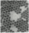
NA is needed for the successful detachment of nascent influenza virus particles from host cells. Shown here is one of the original electron micrographs from the work of Peter Palese et al. (taken with permission from P Palese, Tobita, et al., 1974), showing the aggregation of influenza virus particles in the absence of neuraminidase activity [49].
Figure 5
Figure 5
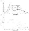
Historic evidence of NA-based protection in human vaccine experiments. (A) Male prisoners vaccinated with X-32 (H7N2) displayed significantly less nasal wash titers when intranasally challenged with H3N2 than those who were vaccinated with influenza B virus. (B) Level of virus in nasal wash specimens was inversely proportional to serum anti-NA antibody titer (taken with permission from Couch et al., 1974) [89].

References

    1. Pappas G., Kiriaze I.J., Falagas M. E. Insights into infectious disease in the era of Hippocrates. Int. J. Infect. Dis. 2008;12:347–350. doi: 10.1016/j.ijid.2007.11.003. - DOI - PubMed
    1. Johnson N.P., Mueller J. Updating the accounts: Global mortality of the 1918–1920 “Spanish” influenza pandemic. Bull. Hist. Med. 2002;76:105–115. doi: 10.1353/bhm.2002.0022. - DOI - PubMed
    1. Wright P.F., Neumann G., Kawaoka Y. Orthomyxoviruses. Fields Virol. 2013;2:1693–1740.
    1. The World Health Organization . Influenza (Seasonal): Fact Sheet No. 211. The World Health Organization; Geneva, Switzerland: 2009.
    1. Molinari N.A., Ortega-Sanchez I.R., Messonnier M.L., Thompson W.W., Wortley P.M., Weintraub E., Bridges C.B. The annual impact of seasonal influenza in the US: Measuring disease burden and costs. Vaccine. 2007;25:5086–5096. doi: 10.1016/j.vaccine.2007.03.046. - DOI - PubMed

Publication types

MeSH terms