. 2014 Jul 16;136(28):9962-72.
doi: 10.1021/ja5016958. Epub 2014 Jul 7.

α-Synuclein-induced membrane remodeling is driven by binding affinity, partition depth, and interleaflet order asymmetry

Affiliations

α-Synuclein-induced membrane remodeling is driven by binding affinity, partition depth, and interleaflet order asymmetry

Anthony R Braun et al. J Am Chem Soc. .

Abstract

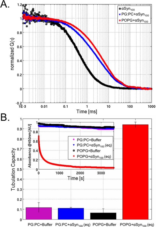
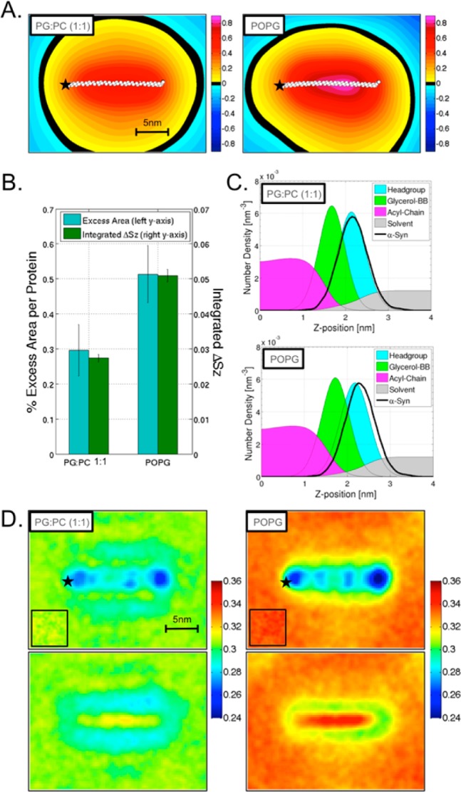
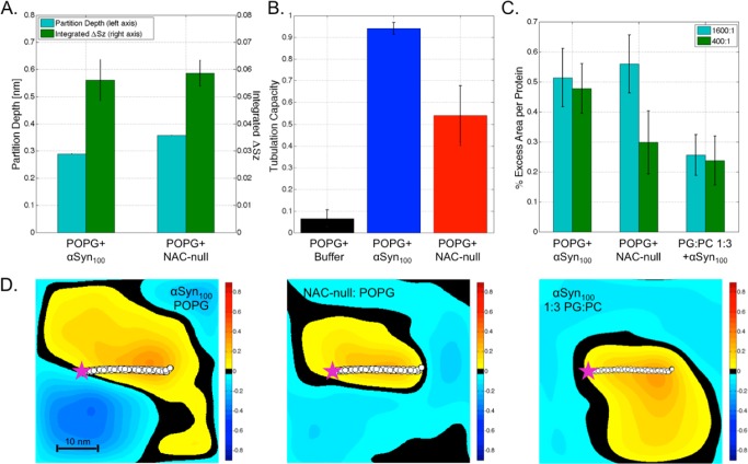
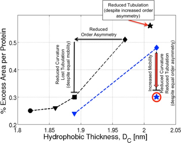
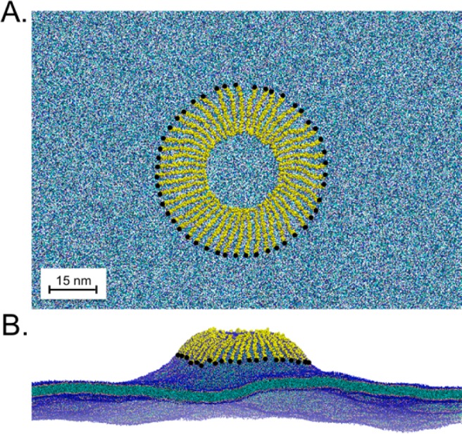
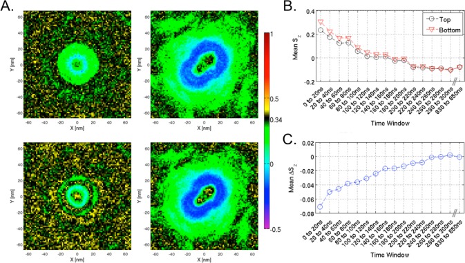
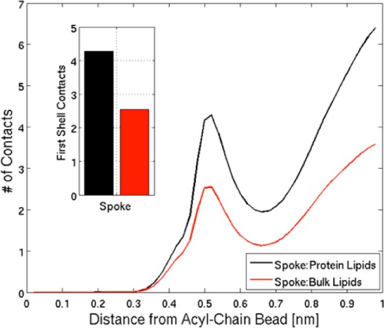
We have investigated the membrane remodeling capacity of the N-terminal membrane-binding domain of α-synuclein (α-Syn100). Using fluorescence correlation spectroscopy and vesicle clearance assays, we show that α-Syn100 fully tubulates POPG vesicles, the first demonstration that the amphipathic helix on its own is capable of this effect. We also show that at equal density of membrane-bound protein, α-Syn has dramatically reduced affinity for, and does not tubulate, vesicles composed of a 1:1 POPG:POPC mixture. Coarse-grained molecular dynamics simulations suggested that the difference between the pure POPG and mixture results may be attributed to differences in the protein's partition depth, the membrane's hydrophobic thickness, and disruption of acyl chain order. To explore the importance of these attributes compared with the role of the reduced binding energy, we created an α-Syn100 variant in which we removed the hydrophobic core of the non-amyloid component (NAC) domain and tested its impact on pure POPG vesicles. We observed a substantial reduction in binding affinity and tubulation, and simulations of the NAC-null protein suggested that the reduced binding energy increases the protein mobility on the bilayer surface, likely impacting the protein's ability to assemble into organized pretubule structures. We also used simulations to explore a potential role for interleaflet coupling as an additional driving force for tubulation. We conclude that symmetry across the leaflets in the tubulated state maximizes the interaction energy of the two leaflets and relieves the strain induced by the hydrophobic void beneath the amphipathic helix.


Figures

Figure 1
Figure 1
(A) FCS traces for α-Syn100 in the absence (black) or presence of equal concentrations of 1:1 PG:PC (blue) or 100% POPG (red) LUVs. The greater shift to the right in the 100% POPG curve reflects a larger fraction of α-Syn100 bound relative to the 1:1 PG:PC curve. (B) α-Syn100 tubulation capacities for 1:1 PG:PC and 100% POPG at equal bound density, compared with buffer. The inset shows the corresponding absorbance traces for systems with 1:1 PG:PC or 100% POPG and buffer or α-Syn100.
Figure 2
Figure 2
(A) Time-averaged height surfaces, ⟨h(x, y)⟩, for 1:1 PG:PC (left) and POPG (right), determined over the last 5 μs of the simulations. Color map units are nm. (B) Percent excess area per protein (blue) and integrated total lipid order (green) for 1:1 PG:PC and POPG. (C) Lipid-component number density profiles for 1:1 PG:PC (top) and POPG (bottom) (solvent, gray; headgroup, cyan; carbonyl-glycerol, green; acyl-chain, magenta; α-Syn100, black line). (D) Local total order parameter Sz(, ) for the membrane near the protein (top) and opposite the protein (bottom) in 1:1 PG:PC (left) and POPG (right) (warm colors = more ordered, cool colors = more disordered). The insets correspond to pure lipid Sz(, ) for each lipid composition. The location of the N-terminus of α-Syn100 is indicated with ★ (the protein itself is not shown).
Figure 3
Figure 3
(A) Comparison of protein partition depths and integrated order parameter asymmetries for α-Syn100 (left) and NAC-null (right) proteins. (B) Experimental tubulation capacities at equal bound protein density for POPG + buffer (black), POPG + α-Syn100 (blue), and POPG + NAC-null (red). (C) Comparison of excess areas per protein for the low-density (1600:1, blue) and high-density (400:1, green) systems for α-Syn100 (left) and NAC-null (middle) in POPG and α-Syn100 in 1:3 PG:PC (right). (D) Time-averaged height surfaces for high-density (400:1) α-Syn100 (left) and NAC-null (middle) systems in POPG and α-Syn100 in 1:3 PG:PC (right). The average protein position is indicated with white spheres, and the N-terminus of the protein is indicated with ★.
Figure 4
Figure 4
Excess area as a function of hydrophobic thickness. Colors demarcate low-density (1600:1, black) vs high-density (400:1, blue). Symbols indicate lipid composition: ● = POPC; ▼ = 1:3 PG:PC; ■ = 1:1 PG:PC; ⧫ = POPG. The ★ denotes NAC-null. Annotations are as described in the text.
Figure 5
Figure 5
(A) Top-down view of the spoke starting configuration. The system includes 48 α-Syn100 proteins (yellow) and 85 296 POPG lipids (blue). Waters have been removed for clarity. The N-terminus of each protein is indicated by ●. (B) Snapshot at 300 ns simulation time. The budding tubule extends ∼25 nm above the bulk lipid bilayer.
Figure 6
Figure 6
(A) Sz(, ) for the protein-containing leaflet (top) and opposite leaflet (bottom) at the early stage (0 to 20 ns, left) and the late tubule stage (830 to 850 ns, right). The color map reference (black) is set at Sz for pure POPG. (B) Time course of the mean Sz for the protein (black) and opposite (red) monolayers as the tubule develops. (C) Time course of the average difference ΔSz across the membrane (ΔSz = Sz,oppositeSz,protein).
Figure 7
Figure 7
Total number of interleaflet contacts (acyl-chain-to-acyl-chain) for lipids near the protein (black) vs lipids in the bulk (red) in the spoke conformation. The number of first-shell contacts is quantified in the inset.

Similar articles

Cited by

References

    1. Robotta M.; Braun P.; van Rooijen B.; Subramaniam V.; Huber M.; Drescher M. ChemPhysChem 2011, 12, 267. - PubMed
    1. Georgieva E. R.; Ramlall T. F.; Borbat P. P.; Freed J. H.; Eliezer D. J. Biol. Chem. 2010, 285, 28261. - PMC - PubMed
    1. Trexler A. J.; Rhoades E. Biochemistry 2009, 48, 2304. - PMC - PubMed
    1. Georgieva E. R.; Ramlall T. F.; Borbat P. P.; Freed J. H.; Eliezer D. J. Am. Chem. Soc. 2008, 130, 12856. - PMC - PubMed
    1. Jao C. C.; Hegde B. G.; Chen J.; Haworth I. S.; Langen R. Proc. Natl. Acad. Sci. U.S.A. 2008, 105, 19666. - PMC - PubMed

Publication types

MeSH terms

LinkOut - more resources