. 2014 Oct;99(10):E1913-21.
doi: 10.1210/jc.2014-1340. Epub 2014 Jun 24.

Dysregulated sphingolipid metabolism in endometriosis

Affiliations

Dysregulated sphingolipid metabolism in endometriosis

Yie Hou Lee et al. J Clin Endocrinol Metab. 2014 Oct.

Abstract

Background: In endometriosis, the establishment and subsistence of ectopic lesions outside the endometrium suggest an altered cellular state for pathological hyperplasia. Sphingolipids are bioactive compounds, and their biosynthesis and metabolism modulate a range of cellular processes including proliferation, migration and apoptosis. We demonstrate that aberrations in sphingolipid metabolism occur in women with endometriosis.

Methods: Targeted mass spectrometry on >120 sphingolipids were measured in the sera (n = 62), peritoneal fluid (n = 63), and endometrial tissue (n = 14) of women with and without endometriosis. Quantitative RT-PCR and immunohistochemistry were performed on endometrial tissues determine the expression levels of sphingolipid enzymes.

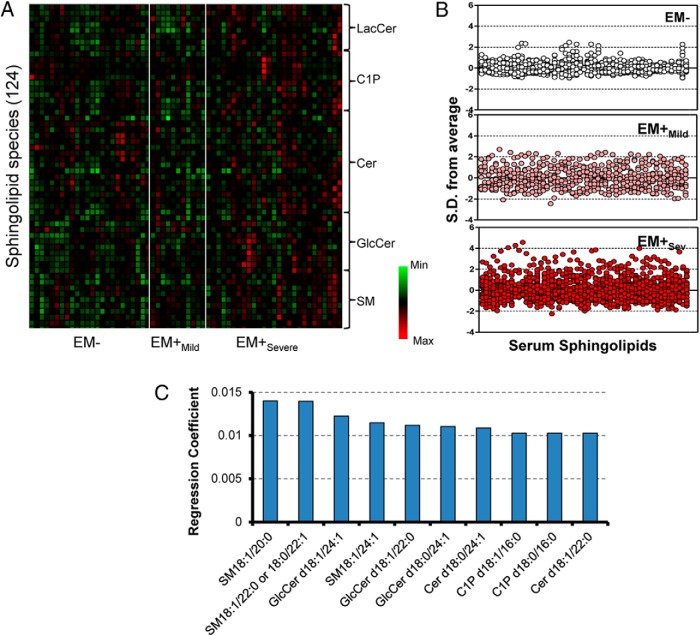
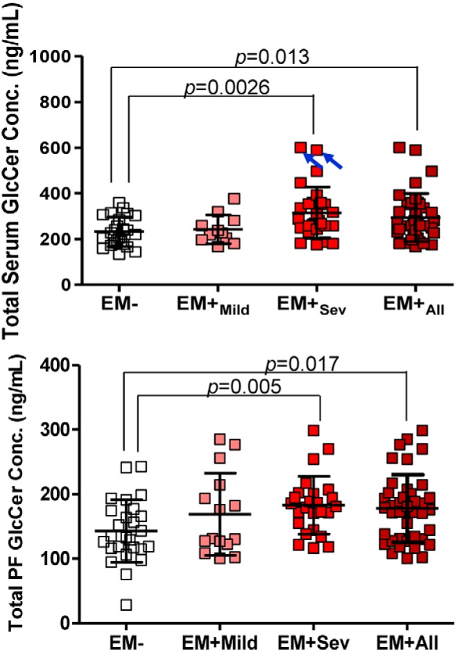
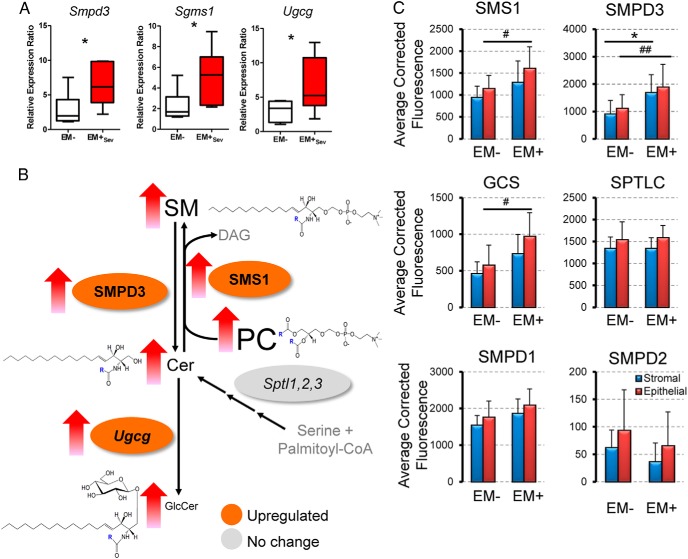
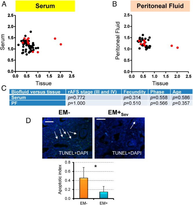
Results: Sphingolipidomics identified the in vivo accumulation of numerous sphingolipids, including the functionally antagonistic glucosylceramides and ceramides in the serum and PF of women with endometriosis. We found upregulation of specific sphingolipid enzymes, namely sphingomyelin synthase 1 (SMS1), sphingomyelinase 3 (SMPD3), and glucosylceramide synthase (GCS) in the endometrium of endometriotic women with corresponding increased GlcCer, decreased sphingomyelin levels, and decreased apoptosis in the endometrium.

Conclusions: Our sphingolipidomics approach provided evidence of altered sphingolipid metabolism flux in serum, peritoneal fluid, and endometrial tissue in women with endometriosis. The results provide new information on how sphingolipids and eutopic endometrium may contribute to the pathophysiology of endometriosis. The results also have implications for the use of sphingolipids as potential biomarkers.


Figures

Figure 1.
Figure 1.. Serum sphingolipid profile of endometriosis.
A, Heat map representation of median-centered, SD-scaled data from women of differential endometriotic stages as determined by rAFS classification (EM, n = 24; EM+Mild, n = 11; EM+Sev, n = 27). Each row represents a sphingolipid species (total 124 species), whereas each column represents a subject grouped together according to their endometriotic status. B, Z-score plots of serum sphingolipids in subjects without endometriosis (EM), mild endometriosis (EM+Mild), severe endometriosis (EM+Sev), and all grades of endometriosis (EM+All). C, Regression coefficients of te top 10 ranked important variables (sphingolipids) from serum that have potential influence on endometriosis.
Figure 2.
Figure 2.. Accumulation of GlcCer in endometriosis.
Scatter plots of total serum and PF GlcCer in EM+ and EM subjects. Open boxes, EM (n = 24); pink boxes, EM+Mild (n = 11); red boxes, EM+Sev (n = 27); maroon boxes, EM+All (n = 38). Although 2 EM+Sev patients seemingly contribute to the difference (blue arrows), their removal maintained statistical significance (P = .007). P values adjusted by Bonferroni adjustment.
Figure 3.
Figure 3.. Regulation of sphingolipid concentrations in endometriosis in the endometrium.
A, Upregulation of endometrial sphingolipid enzymes in EM+Sev (n = 5 per group). *, P < .05. B, Serum lipid concentrations and lipid enzymes that control their metabolism in the endometrium of EM+Sev and EM. Colors indicate fold changes in the expression levels of lipid enzymes that regulate the flux of sphingolipids. Font sizes are proportionate to the abundance of the lipids in serum. Unmeasured analytes are shown in gray. C, Endometrial sphingolipid enzyme expression levels as measured by fluorescence intensity in stromal (blue bar) and epithelial (red bar) compartments (n = 6). *, P < .05 for stromal comparison; and #, P < .05; ##, P < .01 for epithelial comparisons.
Figure 4.
Figure 4.. Accumulation of GlcCer in endometriosis.
A, Scatterplot of mean serum sphingolipid levels against mean endometrial tissue sphingolipid levels. B, Scatter plot of mean PF sphingolipid levels against mean endometrial tissue sphingolipid levels. GlcCers are highlighted in red. C, Because the tissue and PF/serum data were not necessarily from the same patients, Fisher's exact test and Wilcoxon P values of demographics variables in EM+Sev patients were tested and shown to be similar. D, Apoptotic index based on average number of apoptotic bodies/6.4 mm2 detected by TUNEL IHC of EM+ and EM women (n = 4–5). *, P < .05. Original magnification, ×400; scale bar, 50 μm.

References

    1. Practice Committee of the American Society for Reproductive Medicine. Endometriosis and infertility. Fertil Steril. 2004;81:1441–1446. - PubMed
    1. Painter JN, Anderson CA, Nyholt DR, et al. . Genome-wide association study identifies a locus at 7p15.2 associated with endometriosis. Nat Genet. 2011;43:51–54. - PMC - PubMed
    1. Stefansson H, Geirsson RT, Steinthorsdottir V, et al. . Genetic factors contribute to the risk of developing endometriosis. Hum Reprod. 2002;17:555–559. - PubMed
    1. Zondervan KT, Cardon LR, Kennedy SH. The genetic basis of endometriosis. Curr Opin Obstet Gynecol. 2001;13:309–314. - PubMed
    1. Siedentopf F, Tariverdian N, Rücke M, Kentenich H, Arck PC. Immune status, psychosocial distress and reduced quality of life in infertile patients with endometriosis. Am J Reprod Immunol. 2008;60:449–461. - PubMed

Publication types

MeSH terms