. 2014 Sep:74:64-73.
doi: 10.1016/j.freeradbiomed.2014.06.012. Epub 2014 Jun 21.

Arsenic induces sustained impairment of skeletal muscle and muscle progenitor cell ultrastructure and bioenergetics

Affiliations

Arsenic induces sustained impairment of skeletal muscle and muscle progenitor cell ultrastructure and bioenergetics

Fabrisia Ambrosio et al. Free Radic Biol Med. 2014 Sep.

Abstract

Over 4 million individuals in the United States, and over 140 million individuals worldwide, are exposed daily to arsenic-contaminated drinking water. Human exposures can range from below the current limit of 10 μg/L to over 1mg/L, with 100 μg/L promoting disease in a large portion of those exposed. Although increased attention has recently been paid to myopathy following arsenic exposure, the pathogenic mechanisms underlying clinical symptoms remain poorly understood. This study tested the hypothesis that arsenic induces lasting muscle mitochondrial dysfunction and impairs metabolism. Compared to nonexposed controls, mice exposed to drinking water containing 100 μg/L arsenite for 5 weeks demonstrated impaired muscle function, mitochondrial myopathy, and altered oxygen consumption that were concomitant with increased mitochondrial fusion gene transcription. There were no differences in the levels of inorganic arsenic or its monomethyl and dimethyl metabolites between controls and exposed muscles, confirming that arsenic does not accumulate in muscle. Nevertheless, muscle progenitor cells isolated from exposed mice recapitulated the aberrant myofiber phenotype and were more resistant to oxidative stress, generated more reactive oxygen species, and displayed autophagic mitochondrial morphology, compared to cells isolated from nonexposed mice. These pathological changes from a possible maladaptive oxidative stress response provide insight into declines in muscle functioning caused by exposure to this common environmental contaminant.

Keywords: Free radicals; Metabolism; Mitochondria; Resistance to stress.


Figures

Figure 1
Figure 1
Arsenic impairs muscle function. Mice were exposed to 0 (n=6) or 100 µg/L (n=5) As(III) in drinking water for 5 wk. At the end of the exposure, muscle contractile function was tested in situ, as described in methods. Following assessment of peak force production (Table II), animals were subjected to a 7-minute high-frequency fatiguing protocol with 350ms train stimulation at 100Hz every 4 seconds. Force recovery was analyzed at 5 min following completion of the fatiguing protocol. Data are presented as mean + SEM of the % of starting force. Linear regression analysis demonstrated that the slopes of the control and As(III) fatigue curves are significantly different (#; p=0.02). * designates statistical difference from control at p<0.001, as determined by two-way analysis of variance.
Figure 2
Figure 2
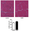
Arsenic exposure results in myofiber hypertrophy. Mice were exposed to 0 or 100 ppb As(III) in drinking water for 5 wk. At the end of exposure, anterior tibialis muscles were isolated, sectioned and stained with H&E. A. shows brightfield 20× images of muscle with increased swollen fibers in the As(III)-exposed mice. B. Myofiber area in 50 fibers in each of three cross-sections was quantified and averaged for each mouse and data are presented as mean ± SEM myofiber area in the different exposure groups (As(III) n=6) versus n=7 control muscles). * designates statistical difference from control at p<0.05, as determined by a standard t-test.
Figure 3
Figure 3
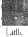
Arsenic induces dysfuctional myofiber mitochondria and bioenergetics. A. Anterior tibialis muscles were fixed and processed for TEM as indicated in methods. The 10000× and 50,000× images demonstrate progressive ultrastuctural increases in myofiber width and mitochondrial size in mice exposed for 5 wk to 100 µg/L As(III), relative to 5 wk control mice. Images shown are representative of images from 6 mice/ group. B. Mitochondrial oxygen consumption was assessed using high-resolution respirometry in, permeablized myofibers, freshly isolated from control (open bar) and As(III)-exposed (grey bar) mice, as described in methods. Additions to the oxygen consumption chamber were: FB: fiber bundles with no substrate, PMG: pyruvate, malate, glutamate, D: ADP, C: cytochrome C, S: succinate, FCCP: (uncoupled complex 1); Rot: rotenone (inhibited complex 1), Ama: antimycin A (inhibited complex 1 and 2). The data in the graph are mean ± SEM pmol/sec of oxygen consumption normalized to mg of tissue dry weight. Significance between consumption rates at each step for fibers from As(III) exposed mice relative to controls at p<0.05 is designated by * (t-test, n=5 mice in each group).
Figure 4
Figure 4
Time dependent As(III)-induced dysfunctional muscle progenitor cell phenotype. Progenitor cells were isolated from hindlimb muscle of mice exposed to 0 or 100 ppb As(III) for 5 wk, as well as mice exposed to 100 µg/L As(III) for 2 wk. (A) Ultrastructural analysis by TEM imaging revealed that cells isolated from 2 and 5 wk As(III)-exposed mice retained the swollen and dysfunctional mitochondrial (M) phenotype. Cells isolated from 5 wk exposed mice indicated that the additional exposure induced a sustained increase of lyosomes (L) and autophagolysosomes (AL). The images are representative of multiple cells in replicate cultures of cells isolated from the different groups. (B). Mitochondrial-driven respiration was compared in progenitor cells isolated from control or 5 wk As(III)-exposed mice after expansion in culture without As(III), as described in methods. Nonphosphorylating LEAK respiration (electon flow coupled to proton pumping to compensate for proton leak) was measured in the presence of oligomycin. The data in the graph are mean ± SEM pmol of oxygen flow/sec per million cells. Significance between consumption rates at each step for fibers from As(III) exposed mice relative to controls of p<0.002 is designated by ** (t-test, n=4).
Figure 5
Figure 5
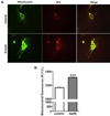
As(III) induces a muscle progenitor cell phenotype with enhanced mitochondrial superoxide generation. Progenitor cells derived from control or As(III)-exposed mice were cultured without As(III) in chamber dishes for live cell confocal imaging of Mitosox™ Red dye oxydation. Cells were loaded with both Mitosox™ and Mito tracker® Green and followed over 30 minutes for change in respective fluoresence intensity. A. shows typical images of control cells and cells from As(III)-exposed mice. B. Fluorecent intensity of Mitosox™ relative to Mito tracker® was quantified and data are plotted as mean + SEM RFU for 2500–3000 cells in each culture. The data are repesentive of three replicate cell isolations and *** designates significant difference at p<0.001.
Figure 6
Figure 6
As(III) induces a muscle progenitor cell phenotype that resists oxidant stress. Progenitor cells isolated from control or 5 wk As(III)-exposed mice were compared for cell number over 72 h following exposure to a toxic oxidative stress from 200µM H2O2. Cells from both groups were exposed to H2O2 at time=0 and cellular numbers were analyzed using Live Automated Cell Imaging. The data are normalized to the number of cells per 10× microscopic field with the same nine locations imaged at each time point in the respective cultures. * designates difference p<0.05 from respective control at the designated time point.

Similar articles

Cited by

References

    1. Chakraborti D, Mukherjee SC, Pati S, Sengupta MK, Rahman MM, Chowdhury UK, Lodh D, Chanda CR, Chakraborti AK, Basu GK. Arsenic groundwater contamination in Middle Ganga Plain, Bihar, India: a future danger? Environ Health Perspect. 2003;111:1194–1201. - PMC - PubMed
    1. Mukherjee SC, Rahman MM, Chowdhury UK, Sengupta MK, Lodh D, Chanda CR, Saha KC, Chakraborti D. Neuropathy in arsenic toxicity from groundwater arsenic contamination in West Bengal, India. J Environ Sci Health A Tox Hazard Subst Environ Eng. 2003;38:165–183. - PubMed
    1. Parvez F, Wasserman GA, Factor-Litvak P, Liu X, Slavkovich V, Siddique AB, Sultana R, Sultana R, Islam T, Levy D, et al. Arsenic exposure and motor function among children in Bangladesh. Environ Health Perspect. 2011;119:1665–1670. - PMC - PubMed
    1. Guha Mazumder DN. Chronic arsenic toxicity: clinical features, epidemiology, and treatment: experience in West Bengal. J.Environ.Sci Health Part A Tox.Hazard.Subst.Environ.Eng. 2003;38:141–163. - PubMed
    1. Doherty TJ. Invited review: Aging and sarcopenia. J Appl Physiol. 2003;95:1717–1727. - PubMed

Publication types

MeSH terms