

Iron excretion in iron dextran-overloaded mice
- PMID: 24960657
- PMCID: PMC4212028
- DOI: 10.2450/2014.0288-13
Iron excretion in iron dextran-overloaded mice
Abstract
Background: Iron homeostasis in humans is tightly regulated by mechanisms aimed to conserve iron for reutilisation, with a negligible role played by excretory mechanisms. In a previous study we found that mice have an astonishing ability to tolerate very high doses of parenterally administered iron dextran. Whether this ability is linked to the existence of an excretory pathway remains to be ascertained.
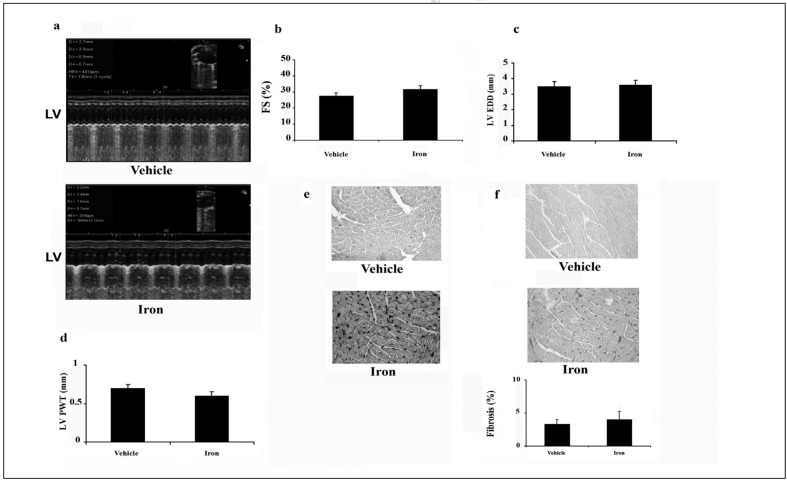
Materials and methods: Iron overload was generated by intraperitoneal injections of iron dextran (1 g/kg) administered once a week for 8 weeks in two different mouse strains (C57bl/6 and B6D2F1). Urinary and faecal iron excretion was assessed by inductively coupling plasma-mass spectrometry, whereas cardiac and liver architecture was evaluated by echocardiography and histological methods. For both strains, 24-hour faeces and urine samples were collected and iron concentration was determined on days 0, 1 and 2 after iron administration.
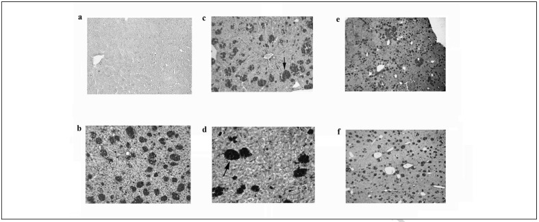
Results: In iron-overloaded C57bl/6 mice, the faecal iron concentration increased by 218% and 157% on days 1 and 2, respectively (p<0.01). The iron excreted represented a loss of 14% of total iron administered. Similar but smaller changes was also found in B6D2F1 mice. Conversely, we found no significant changes in the concentration of iron in the urine in either of the strains of mice. In both strains, histological examination showed accumulation of iron in the liver and heart which tended to decrease over time.
Conclusions: This study indicates that mice have a mechanism for removal of excess body iron and provides insights into the possible mechanisms of excretion.
Figures
References
-
- Ganz T, Nemeth E. Iron imports. IV. Hepcidin and regulation of body iron metabolism. Am J Physiol. 2006;290:G199–G203. - PubMed
-
- Porter JB. Practical management of iron overload. Br J Haematol. 2001;115:239–52. - PubMed
-
- Borgna-Pignatti C, Rugolotto S, De Stefano P, et al. Survival and complications in patients with thalassemia major treated with transfusion and deferoxamine. Haematologica. 2004;89:1187–93. - PubMed
-
- Bonkovsky HL, Healey JF, Lincoln B, et al. Hepatic heme synthesis in a new model of experimental hemochromatosis: studies in rats fed finely divided elemental iron. Hepatology. 1987;7:1195–203. - PubMed
Publication types
MeSH terms
Substances
LinkOut - more resources
Full Text Sources
Medical
