

Multiple thoracolithiasis: An incidental finding
- PMID: 24960759
- PMCID: PMC3649582
- DOI: 10.1093/jscr/2012.8.1
Multiple thoracolithiasis: An incidental finding
Abstract
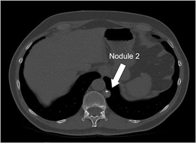
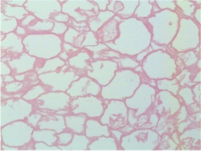
Thoracoliths are rare benign intrapleural fibrotic structures with a necrotic fat core. There are 19 previous reported cases in the literature. This case report presents for the first time, a patient with two thoracoliths within the same hemithorax. Both lesions were identified incidentally in the left hemithorax by computed tomography and remained in the same position on repeat imaging. The lesions were removed by a video-assisted thoracic surgery approach. Histology revealed a 20mm and a 14mm lesion, with a fibrotic dense collagen shell surrounding a non-viable necrotic fat core. This case demonstrates that thoracolithiasis is a rare differential diagnosis for incidental multiple non-mobile lesions within the thorax.
© JSCR.
Figures


References
-
- Dias AR, Zerbini EJ, Curi N. Pleural stone. A case report. The Journal of thoracic and cardiovascular surgery. 1968;56(1):120–2 - PubMed
-
- Iwasaki T, Nakagawa K, Katsura H, Ohse N, Nagano T, Kawahara K. Surgically removed thoracolithiasis: report of two cases. Annals of thoracic and cardiovascular surgery: official journal of the Association of Thoracic and Cardiovascular Surgeons of Asia. 2006;12(4):279–82 - PubMed
-
- Kosaka S, Kondo N, Sakaguchi H, Kitano T, Harada T, Nakayama K. Thoracolithiasis. The Japanese journal of thoracic and cardiovascular surgery : official publication of the Japanese Association for Thoracic Surgery = Nihon Kyobu Geka Gakkai zasshi. 2000;48(5):318–21 - PubMed
-
- Bolca C, Trahan S, Frechette E. Intrapleural thoracolithiasis: a rare intrathoracic pearl-like lesion. The Thoracic and cardiovascular surgeon. 2011;59(7):445–6 - PubMed
-
- Chujo M, Miura T, Kawano Y, Miyawaki M, Imakiire T, Hayashita Y, et al. [Thoracolithiasis detected during video-assisted thoracoscopic surgery for spontaneous pneumothorax: report of a case]. Kyobu geka The Japanese journal of thoracic surgery. 2005;58(13):1173–6 - PubMed
LinkOut - more resources
Full Text Sources
