

Treatment of MSCs with Wnt1a-conditioned medium activates DP cells and promotes hair follicle regrowth
- PMID: 24961246
- PMCID: PMC4069670
- DOI: 10.1038/srep05432
Treatment of MSCs with Wnt1a-conditioned medium activates DP cells and promotes hair follicle regrowth
Abstract
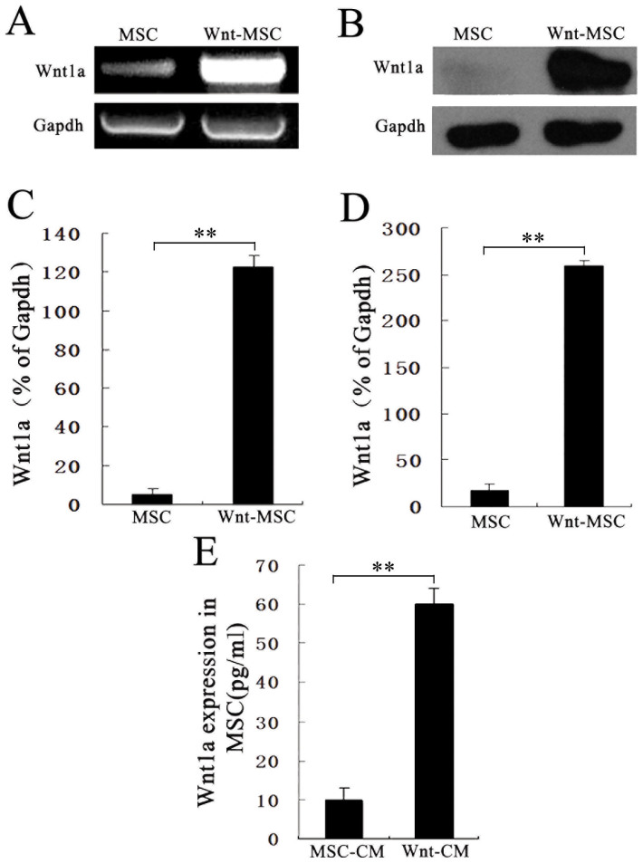
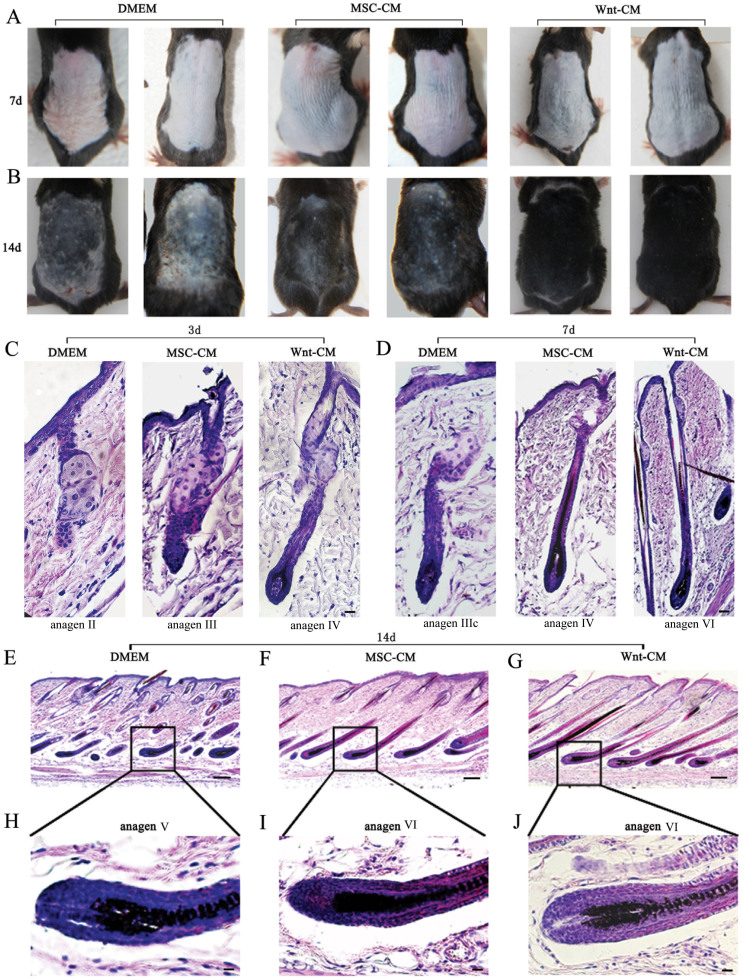
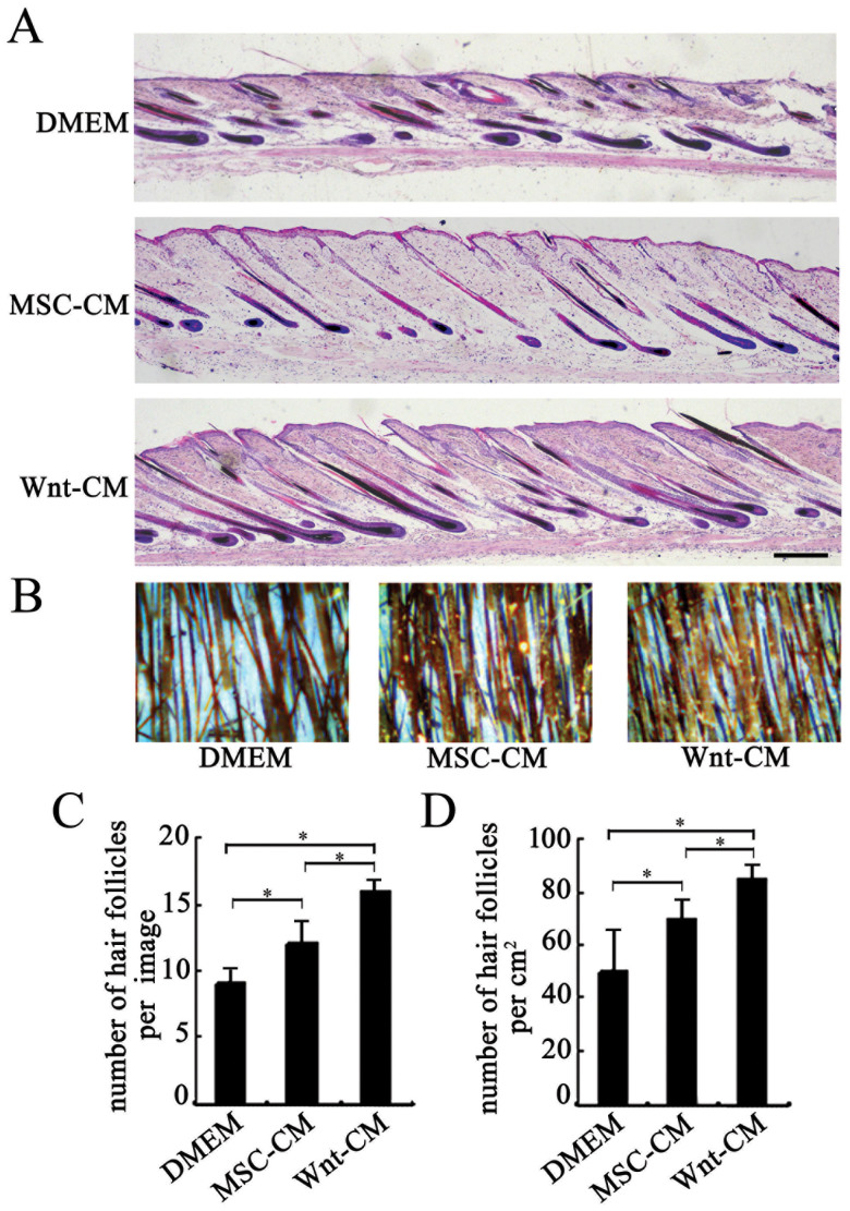
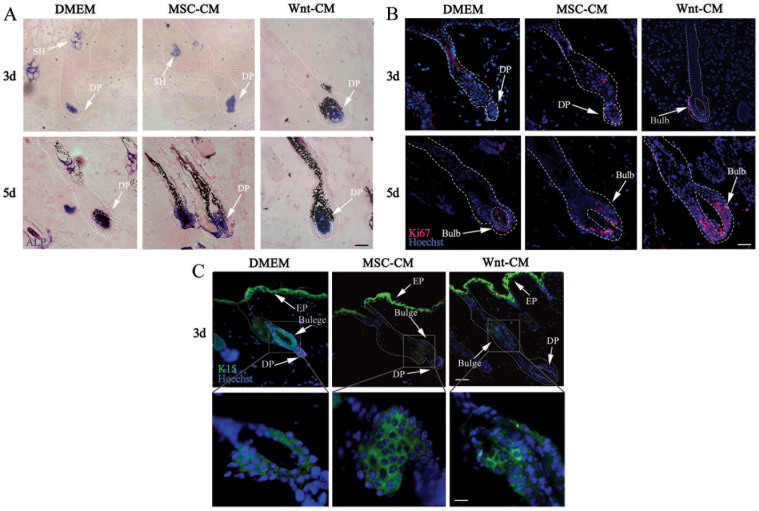
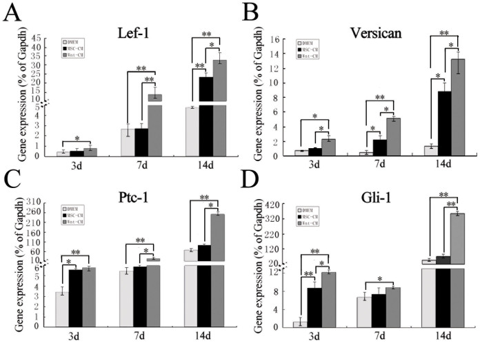
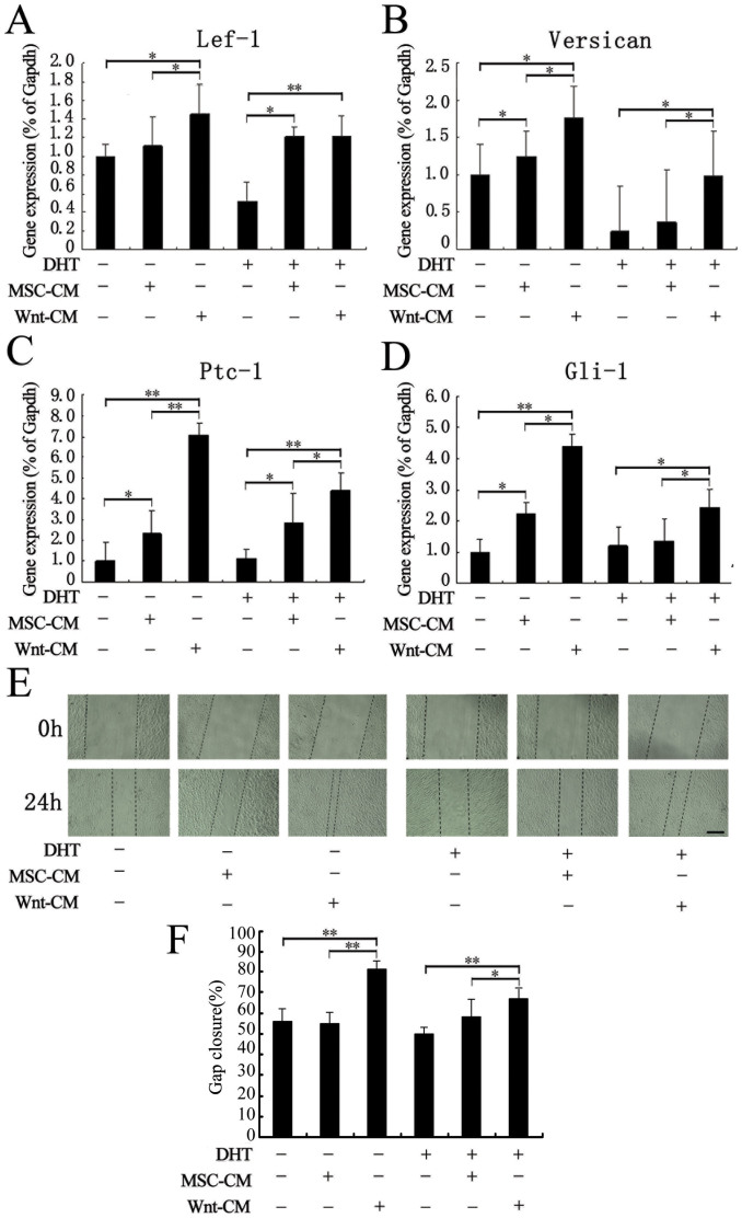
Hair loss (alopecia) is a common problem for people. The dermal papilla is the key signaling center that regulates hair growth and it engage in crosstalk with the microenvironment, including Wnt signaling and stem cells. In this study, we explored the effects of bone marrow mesenchymal stem cell overexpression of Wnt1a on mouse hair follicle regeneration. Wnt-CM accelerated hair follicle progression from telogen to anagen and enhanced the ALP expression in the DP area. Moreover, the hair induction-related genes were upregulated, as demonstrated by qRT-PCR. Wnt-CM treatment restored and increased DP cell expression of genes downregulated by dihydrotestosterone treatment, as demonstrated by qRT-PCR assays. Our study reveals that BM-MSC-generated Wnt1a promotes the DP's ability to induce hair cycling and regeneration.
Figures
Similar articles
-
Wnt1a maintains characteristics of dermal papilla cells that induce mouse hair regeneration in a 3D preculture system.J Tissue Eng Regen Med. 2017 May;11(5):1479-1489. doi: 10.1002/term.2046. Epub 2015 Jun 29. J Tissue Eng Regen Med. 2017. PMID: 26118627
-
Overexpression of Nanog in amniotic fluid-derived mesenchymal stem cells accelerates dermal papilla cell activity and promotes hair follicle regeneration.Exp Mol Med. 2019 Jul 4;51(7):1-15. doi: 10.1038/s12276-019-0266-7. Exp Mol Med. 2019. PMID: 31273189 Free PMC article.
-
Wnt activator CHIR99021-stimulated human dermal papilla spheroids contribute to hair follicle formation and production of reconstituted follicle-enriched human skin.Biochem Biophys Res Commun. 2019 Aug 27;516(3):599-605. doi: 10.1016/j.bbrc.2019.06.038. Epub 2019 Jun 18. Biochem Biophys Res Commun. 2019. PMID: 31221480
-
Advances in Regenerative Stem Cell Therapy in Androgenic Alopecia and Hair Loss: Wnt pathway, Growth-Factor, and Mesenchymal Stem Cell Signaling Impact Analysis on Cell Growth and Hair Follicle Development.Cells. 2019 May 16;8(5):466. doi: 10.3390/cells8050466. Cells. 2019. PMID: 31100937 Free PMC article. Review.
-
Maintaining Hair Inductivity in Human Dermal Papilla Cells: A Review of Effective Methods.Skin Pharmacol Physiol. 2020;33(5):280-292. doi: 10.1159/000510152. Epub 2020 Oct 14. Skin Pharmacol Physiol. 2020. PMID: 33053562 Review.
Cited by
-
Comparative characterization and osteogenic / adipogenic differentiation of mesenchymal stem cells derived from male rat hair follicles and bone marrow.Cell Regen. 2020 Aug 11;9(1):13. doi: 10.1186/s13619-020-00051-7. Cell Regen. 2020. PMID: 32778979 Free PMC article.
-
H3K4me3 regulates the transcription of RSPO3 in dermal papilla cells to influence hair follicle morphogenesis and development.Epigenetics Chromatin. 2025 Aug 8;18(1):52. doi: 10.1186/s13072-025-00611-8. Epigenetics Chromatin. 2025. PMID: 40775776 Free PMC article.
-
Anti-CXCL4 monoclonal antibody accelerates telogen to anagen transition and attenuates apoptosis of the hair follicle in mice.Exp Ther Med. 2017 Aug;14(2):1001-1008. doi: 10.3892/etm.2017.4578. Epub 2017 Jun 12. Exp Ther Med. 2017. PMID: 28810552 Free PMC article.
-
A Conditioned Medium of Umbilical Cord Mesenchymal Stem Cells Overexpressing Wnt7a Promotes Wound Repair and Regeneration of Hair Follicles in Mice.Stem Cells Int. 2017;2017:3738071. doi: 10.1155/2017/3738071. Epub 2017 Feb 27. Stem Cells Int. 2017. PMID: 28337222 Free PMC article.
-
The Effect of Mesenchymal Stem Cells Derived-Conditioned Media in Combination with Oral Anti-Androgenic Drugs on Male Pattern Baldness: An Animal Study.Cell J. 2023 Nov 1;25(11):790-800. doi: 10.22074/cellj.2023.2008138.1377. Cell J. 2023. PMID: 38071411 Free PMC article.
References
-
- Andl T., Reddy S. T., Gaddapara T. & Millar S. E. WNT signals are required for the initiation of hair follicle development (2002). - PubMed
-
- Mc Elwee K. J., Kissling S., Wenzel E., Huth A. & Hoffmann R. Cultured peribulbar dermal sheath cells can induce hair follicle development and contribute to the dermal sheath and dermal papilla. J Invest Dermatol 121, 1267–1275 (2003). - PubMed
Publication types
MeSH terms
Substances
LinkOut - more resources
Full Text Sources
Other Literature Sources
