. 2015 Dec;16(4):1135-48.
doi: 10.1177/1470320313515039. Epub 2014 Jun 24.

Nuclear expression of renin-angiotensin system components in NRK-52E renal epithelial cells

Affiliations

Nuclear expression of renin-angiotensin system components in NRK-52E renal epithelial cells

Ebaa M Alzayadneh et al. J Renin Angiotensin Aldosterone Syst. 2015 Dec.

Abstract

Introduction: Isolated nuclei of sheep proximal tubules express angiotensin (Ang) receptors as well as angiotensinogen (AGT) and renin. The present study characterized the NRK-52E tubular epithelial cell line for the intracellular expression of renin-angiotensin system (RAS) components.

Methods: RAS components were visualized by immunofluorescent staining in intact cells and protein expression in isolated nuclei.

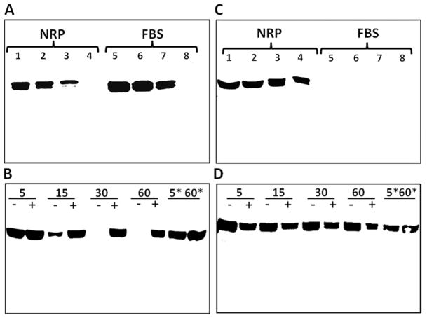
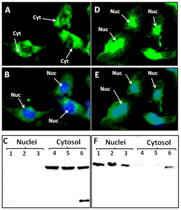
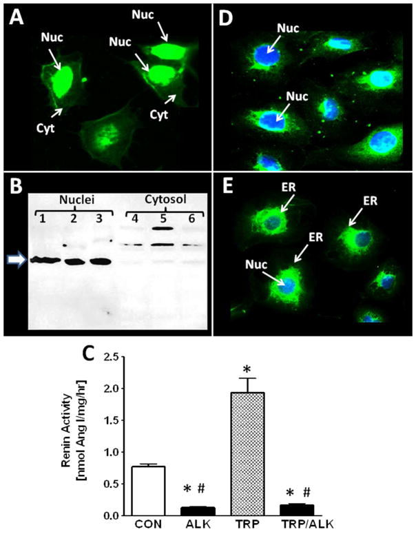
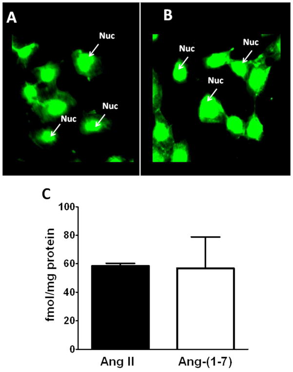
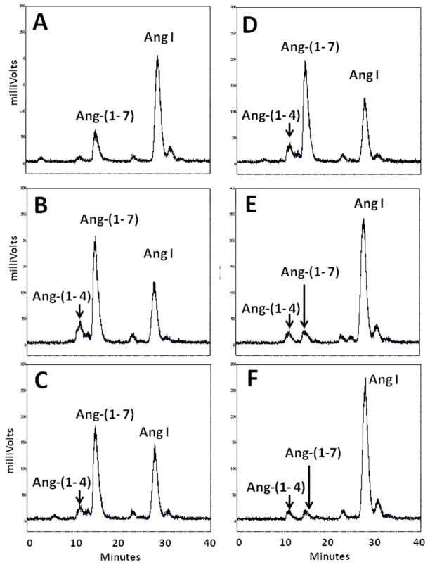
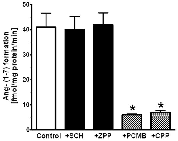
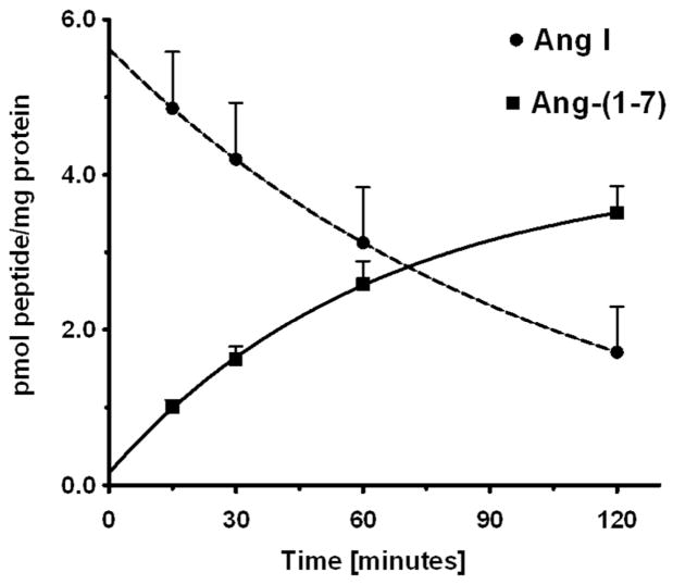
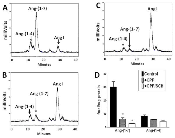
Results: An antibody to the angiotensin I (Ang I) sequence of AGT (AI-AGT) revealed only cytosolic staining, while an antibody to an internal sequence of AGT (Int-AGT) revealed primarily nuclear staining. Immunoblots of nuclear and cytosolic fractions confirmed the differential cell staining of AGT. Immunostaining for renin was present on nuclei of intact cells. Nuclear renin activity averaged 0.77±0.05 nmol/mg protein/h that was reduced by aliskiren (0.13±0.01 nmol/mg/h, n=3, p<0.01); trypsin activation increased activity three-fold. Peptide staining localized angiotensin II (Ang II) and Ang-(1-7) to the nucleus and peptide content averaged 59±2 and 57±22 fmol/mg (n=4), respectively. Peptide metabolism in isolated nuclei revealed the processing of Ang I to Ang-(1-7) by thimet oligopeptidase.

Conclusion: We conclude that the NRK-52E cells express an intracellular RAS localized to the nucleus and may be an appropriate cell model to elucidate the functional relevance of this system.

Keywords: NRK-52E; angiotensin; angiotensinogen; renin; thimet oligopeptidase.


Conflict of interest statement

CONFLICT OF INTERESTS

None declared

Figures

Figure 1
Figure 1
Characterization of antibodies to rat and bovine angiotensinogen (AGT). Panel A: Immunoblot of nephrectomized rat plasma (NRP) and fetal bovine serum (FBS) probed with Ang I-AGT (AI-AGT) antibody. The AI-AGT antibody detected AGT bands at 55 and 60 kDa in both NRP and FBS samples. Panel B: incubation of renin with AGT in NRP for 5 to 60 minutes at 37°C or on ice (*5 & *60 minutes) in the absence (−) or presence (+) of the renin inhibitor aliskerin (10 μM) and probed with AI-AGT antibody. Panel C: Immunoblot of NRP and FBS samples with the AGT antibody directed to internal sequence distal to Ang I (Int-AGT) that recognized rat but not bovine AGT. Panel D: the immunoblot from B was stripped and reprobed with the Int-AGT antibody revealing no effect of renin exposure on AGT expression.
Figure 2
Figure 2
Angiotensinogen (AGT) expression in the NRK-52E cells. Panel A: Immunofluorescent (IMF) staining of NRK-52E cells with the Ang I-intact AGT (AI-AGT) antibody reveals predominantly cytosolic (Cyt) staining. Panel B: Identical image of panel A with DAPI nuclear (Nuc) staining in blue. Panel C: Immunoblot of nuclear and cytosolic fractions with AI-AGT antibody reveals a predominant 55 kDa band in cytosolic fractions. Panel D: IMF staining with Int-AGT antibody reveals nuclear associated staining. Panel E: Identical image of panel D with DAPI nuclear (Nuc) staining in blue. Panel F: Immunoblot of nuclear and cytosolic fractions reveals predominant 55 kDa band in the nuclear fraction. IMF staining was representative of 3 different cell passages. Immunoblot fractions were from 3 different cell passages.
Figure 3
Figure 3
Renin and prorenin receptor (PRR) expression in NRK-52E cells. Panel A: Immunofluorescent (IMF) staining of NRK-52E cells with the renin antibody reveals nuclear-associated staining. Panel B: Immunoblot of nuclear and cytosolic fractions with renin antibody reveals a 55 kDa (arrow) band in the nuclear fraction from cells of 3 different passages. Higher molecular weight bands were evident in the cytosolic fractions. Panel C: Renin activity in the nuclear fraction of control (CON) or trypsin-treated (TRP) nuclear lysates treated with or without the renin inhibitor aliskerin (ALK). Values are means ± SEM, *p< 0.01 vs. CON, n = 3 different passages. Panel D: IMF staining of PRR reveals perinuclear staining and DAPI nuclear staining (Nuc) in blue. Panel E: IMF staining of the endoplasmic reticulum (ER) marker calnexin and DAPI nuclear staining (Nuc) in blue.
Figure 4
Figure 4
Expression of Ang II and Ang-(–7) in NRK-52E cells. Immunofluorescent staining using affinity-purified antibodies for Ang II (A) and Ang-(–7) (B) reveal nuclear-associated staining. Panel C: Immunoreactive concentrations of Ang II and Ang-(–7) in the nuclear fractions of NRK-52E cells. Values are means ± SEM; n=4.
Figure 5
Figure 5
Conversion of Ang I to Ang-(–7) in isolated nuclei from NRK-52E cells. HPLC analysis of 125I-Ang I metabolism at 37°C revealed primarily Ang-(–7) (Ang 7) at 15 minutes (A) and 60 minutes (B). The neprilysin inhibitor SCH39370 (SCH; 10 μM) or prolyl oligopeptidase inhibitor z-prolyl-prolinal (ZPP; 50 μM) did not inhibit Ang-(–7) generation (C and D, respectively). The thimet oligopeptidase inhibitor CPP (50 μM) and the thiol inhibitor PCMB (500 μM) essentially abolished Ang-(–7) production (E and F, respectively). A minor peak of Ang-(–4) (Ang 4) was detected in panels B–F.
Figure 6
Figure 6
Influence of peptidase inhibitors on the generation of Ang-(–7) from Ang I in the isolated nuclear fraction of NRK-52E cells. Peptidase inhibitors were neprilysin inhibitor SCH, prolyl oligopeptidase inhibitor ZPP, thimet oligopeptidase inhibitor CPP and thiol protease inhibitor PCMB. All values represent mean ± SEM, n=4 from different cell passages; P < 0.01 versus Control (CON).
Figure 7
Figure 7
Time course for the disappearance of Ang I and formation of Ang-(–7) in isolated nuclei from NRK-52E cells. The conversion of 125I-Ang I to 125I-Ang-(–7) in nuclei at 37°C was quantitated by HPLC separation coupled to a γ-counter. Values are means ± SEM, n=4 of different cell passages. Non-linear decay curve for Ang I and exponential one-phase association of Ang-(–7) were constructed in GraphPad Prism V.
Figure 8
Figure 8
Influence of combined thimet oligopeptidase and neprilysin inhibition on Ang-(–7) and Ang-(–4) formation in isolated nuclei NRK-52E cells. Panel A: Metabolism of 125I-Ang I to Ang-(–7) and Ang-(–4) following 120 minute incubation with nuclei at 37°C. Panel B: Addition of thimet oligopeptidase inhibitor CPP (50 ZM) blocked the majority of Ang-(–7) formation. Panel C: Addition of CPP and neprilysin inhibitor SCH39370 (SCH, 10 ZM). Panel D: Influence of CPP or CPP/SCH on the formation of Ang-(–7) and Ang-(–4) in nuclei at 120 minutes. All values represent mean ± SEM, n=3 from different cell passages; *P < 0.05 versus Control.

Similar articles

Cited by

References

    1. Kobori H, Nangaku M, Navar LG, Nishiyama A. The intrarenal renin-angiotensin system: from physiology to the pathobiology of hypertension and kidney disease. Pharmacol Rev. 2007;59(3):251–87. - PubMed
    1. Paul M, Mehr AP, Kreutz R. Physiology of local renin-angiotensin systems. Physiol Rev. 2006;86:747–803. - PubMed
    1. Velez JC. The importance of the intrarenal renin-angiotensin system. Nat Clin Pract Nephrol. 2009;5(2):89–100. - PubMed
    1. Singh VP, Baker KM, Kumar R. Activation of the intracellular renin-angiotensin system in cardiac fibroblasts by high glucose: role in extracellular matrix production. Am J Physiol Heart Circ Physiol. 2008;294(4):H1675–1684. - PubMed
    1. Singh VP, Le B, Bhat VB, Baker KM, Kumar R. High-glucose-induced regulation of intracellular ANG II synthesis and nuclear redistribution in cardiac myocytes. Am J Physiol Heart Circ Physiol. 2007;293(2):H939–H948. - PubMed

Publication types

MeSH terms

LinkOut - more resources