. 2013 Jun 25;3(3):1013-22.
doi: 10.3390/brainsci3031013.

Neuroprotective role of nerve growth factor in hypoxic-ischemic brain injury

Affiliations

Neuroprotective role of nerve growth factor in hypoxic-ischemic brain injury

Claudia Fantacci et al. Brain Sci. .

Abstract

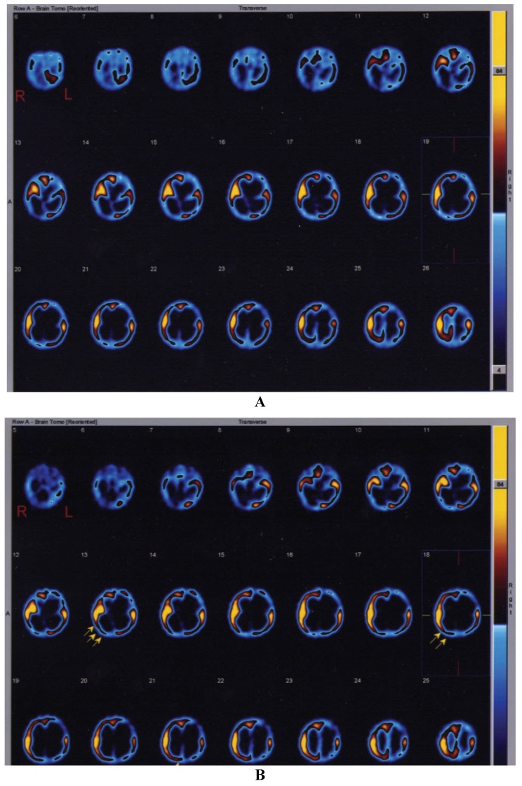
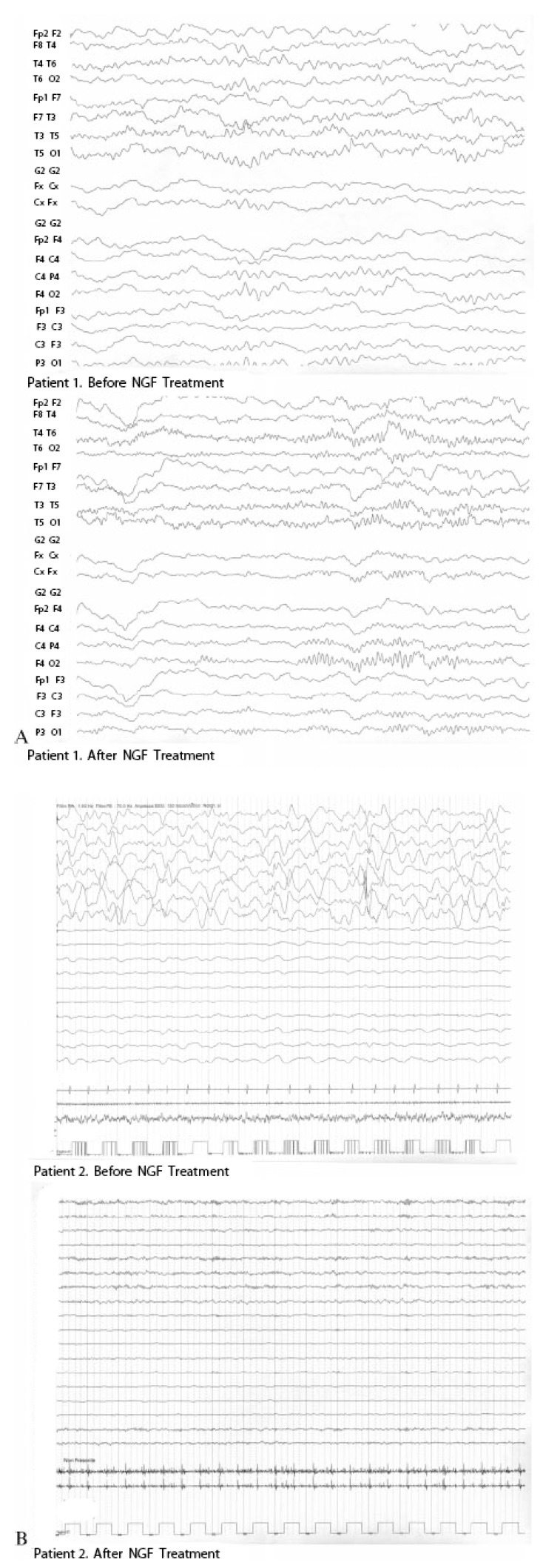
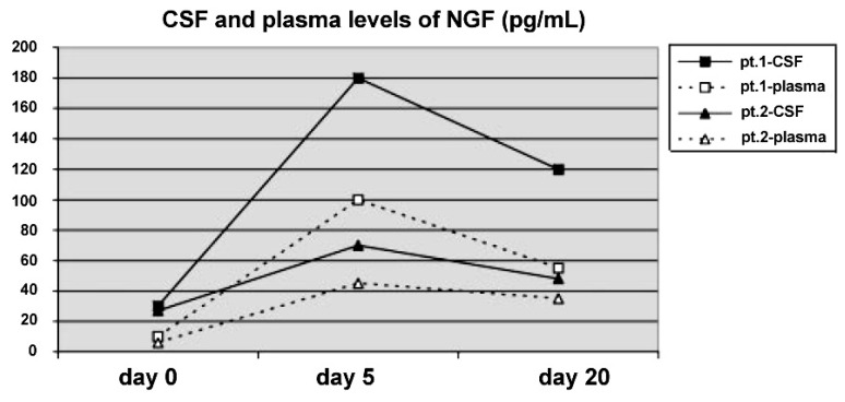
Hypoxic-ischemic brain injuries (HIBI) in childhood are frequently associated with poor clinical and neurological outcome. Unfortunately, there is currently no effective therapy to restore neuronal loss and to determine substantial clinical improvement. Several neurotrophins, such as Nerve Growth Factor (NGF), Brain-Derived Neurotrophic Factor (BDNF), and Glial Derived Neurotrophic Factor (GDNF), play a key role in the development, differentiation, and survival of the neurons of the peripheral and central nervous system. Experimental animal studies demonstrated their neuroprotective role in HIBI, while only a few studies examined the neuroprotective mechanisms in patients with severe HIBI. We report two cases of children with HIBI and prolonged comatose state who showed a significant improvement after intraventricular NGF administration characterized by amelioration of electroencephalogram (EEG) and cerebral perfusion at single-photon emission computed tomography (SPECT). The improvement in motor and cognitive functions of these children could be related to the neuroprotective role exerted by NGF in residual viable cholinergic neurons, leading to the restoration of neuronal networks in the damaged brain.


Figures

Figure 1
Figure 1
Single-photon emission computed tomography (SPECT) study showing the brain perfusion pattern in the second infant affected by severe hypoxic-ischemic injury. (A) Before treatment. (B) After intraventricular NGF infusion. The arrows in (B) highlight the sites of improvement of cerebral perfusion in right temporal and occipital cortices.
Figure 2
Figure 2
Serial electroencephalogram (EEG) examinations performed before and after the treatment with intraventricular Nerve Growth Factor (NGF) administration showed a constant and progressive reduction in slow-wave activity expressed as an increased alpha/theta ratio in both patients (A) patient 1; (B) patient 2.
Figure 3
Figure 3
Cerebrospinal fluid (CSF) and plasma levels of NGF (pg/mL) before, during and after the 10 day course of intraventricular NGF infusion in the two infants. The detection limit was 3 pg/mL.

Similar articles

Cited by

References

    1. Vanucci R.C. Experimental biology of cerebral hypoxia-ischemia: Relation to perinatal brain damage. Pediatr. Res. 1990;27:317–326. doi: 10.1203/00006450-199004000-00001. - DOI - PubMed
    1. Lykissas M.G., Batistatou A.K., Charalabopoulos K.A., Beris A.E. The role of neurotrophins in axonal growth, guidance, and regeneration. Curr. Neurovasc. Res. 2007;4:143–151. doi: 10.2174/156720207780637216. - DOI - PubMed
    1. Han B.H., Holtzman D.M. BDNF protects the neonatal brain from hypoxic-ischemic injury in vivo via the ERK pathway. J. Neurosci. 2000;20:5775–5781. - PMC - PubMed
    1. Wang Y., Chang C.F., Morales M., Chiang Y.H., Hoffer J. Protective effects of glial cell line-derived neurotrophic factor in ischemic brain injury. Ann. N. Y. Acad. Sci. 2002;962:423–437. doi: 10.1111/j.1749-6632.2002.tb04086.x. - DOI - PubMed
    1. Barde Y.A. Neurotrophins: A family of proteins supporting the survival of neurons. Prog. Clin. Biol. Res. 1994;390:1855–1859. - PubMed

LinkOut - more resources