

Effects of Ethanol Exposure during Distinct Periods of Brain Development on Hippocampal Synaptic Plasticity
- PMID: 24961522
- PMCID: PMC4061886
- DOI: 10.3390/brainsci3031076
Effects of Ethanol Exposure during Distinct Periods of Brain Development on Hippocampal Synaptic Plasticity
Abstract
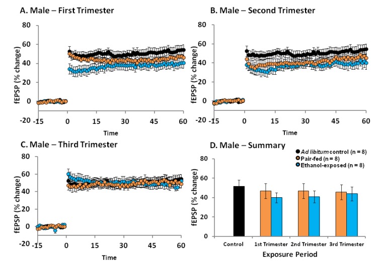
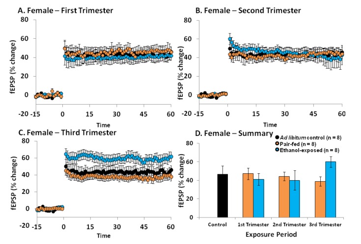
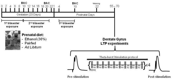
Fetal alcohol spectrum disorders occur when a mother drinks during pregnancy and can greatly influence synaptic plasticity and cognition in the offspring. In this study we determined whether there are periods during brain development that are more susceptible to the effects of ethanol exposure on hippocampal synaptic plasticity. In particular, we evaluated how the ability to elicit long-term potentiation (LTP) in the hippocampal dentate gyrus (DG) was affected in young adult rats that were exposed to ethanol during either the 1st, 2nd, or 3rd trimester equivalent. As expected, the effects of ethanol on young adult DG LTP were less severe when exposure was limited to a particular trimester equivalent when compared to exposure throughout gestation. In males, ethanol exposure during the 1st, 2nd or 3rd trimester equivalent did not significantly reduce LTP in the DG. In females, ethanol exposure during either the 1st or 2nd trimester equivalents did not impact LTP in early adulthood, but following exposure during the 3rd trimester equivalent alone, LTP was significantly increased in the female DG. These results further exemplify the disparate effects between the ability to elicit LTP in the male and female brain following perinatal ethanol exposure (PNEE).
Figures
Similar articles
-
Postnatal Choline Supplementation Rescues Deficits in Synaptic Plasticity Following Prenatal Ethanol Exposure.Nutrients. 2022 May 10;14(10):2004. doi: 10.3390/nu14102004. Nutrients. 2022. PMID: 35631142 Free PMC article.
-
Impaired Bidirectional Synaptic Plasticity in Juvenile Offspring Following Prenatal Ethanol Exposure.Alcohol Clin Exp Res. 2019 Oct;43(10):2153-2166. doi: 10.1111/acer.14170. Epub 2019 Aug 26. Alcohol Clin Exp Res. 2019. PMID: 31386206 Free PMC article.
-
Prenatal ethanol exposure has sex-specific effects on hippocampal long-term potentiation.Hippocampus. 2014 Jan;24(1):54-64. doi: 10.1002/hipo.22203. Epub 2013 Oct 4. Hippocampus. 2014. PMID: 23996604
-
Prenatal ethanol exposure enhances NMDAR-dependent long-term potentiation in the adolescent female dentate gyrus.Hippocampus. 2012 Jan;22(1):69-81. doi: 10.1002/hipo.20849. Epub 2010 Nov 15. Hippocampus. 2012. PMID: 21080406
-
Impairments in hippocampal synaptic plasticity following prenatal ethanol exposure are dependent on glutathione levels.Hippocampus. 2013 Dec;23(12):1463-75. doi: 10.1002/hipo.22199. Epub 2013 Oct 1. Hippocampus. 2013. PMID: 23996467
Cited by
-
A comparison of the different animal models of fetal alcohol spectrum disorders and their use in studying complex behaviors.Front Pediatr. 2014 Sep 3;2:93. doi: 10.3389/fped.2014.00093. eCollection 2014. Front Pediatr. 2014. PMID: 25232537 Free PMC article. Review.
-
Adult Hippocampal Neurogenesis as a Therapeutic Target in Fetal Alcohol Spectrum Disorder.Adv Exp Med Biol. 2025;1473:93-109. doi: 10.1007/978-3-031-81908-7_5. Adv Exp Med Biol. 2025. PMID: 40128476 Review.
-
Effects of prenatal alcohol exposure (PAE): insights into FASD using mouse models of PAE.Biochem Cell Biol. 2018 Apr;96(2):131-147. doi: 10.1139/bcb-2017-0280. Epub 2018 Jan 25. Biochem Cell Biol. 2018. PMID: 29370535 Free PMC article. Review.
-
The effects of developmental alcohol exposure on the neurobiology of spatial processing.Neurosci Biobehav Rev. 2019 Dec;107:775-794. doi: 10.1016/j.neubiorev.2019.09.018. Epub 2019 Sep 14. Neurosci Biobehav Rev. 2019. PMID: 31526818 Free PMC article. Review.
-
Postnatal Choline Supplementation Rescues Deficits in Synaptic Plasticity Following Prenatal Ethanol Exposure.Nutrients. 2022 May 10;14(10):2004. doi: 10.3390/nu14102004. Nutrients. 2022. PMID: 35631142 Free PMC article.
References
-
- Jones K.L. The fetal alcohol syndrome. Addict. Dis. 1975;2:79–88. - PubMed
LinkOut - more resources
Full Text Sources
Other Literature Sources
Research Materials
