. 2013 Nov 12;3(4):1461-82.
doi: 10.3390/brainsci3041461.

White matter loss in a mouse model of periventricular leukomalacia is rescued by trophic factors

Affiliations

White matter loss in a mouse model of periventricular leukomalacia is rescued by trophic factors

Araceli Espinosa-Jeffrey et al. Brain Sci. .

Abstract

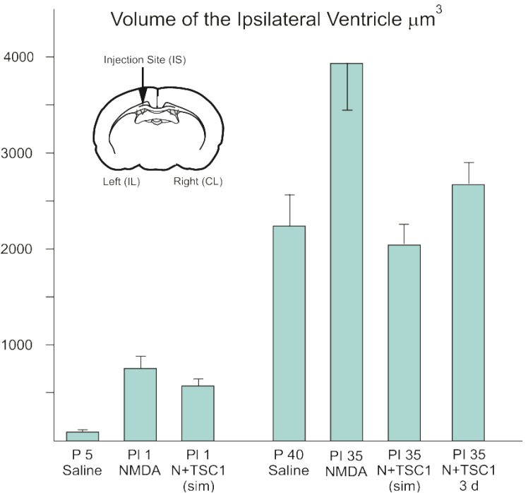
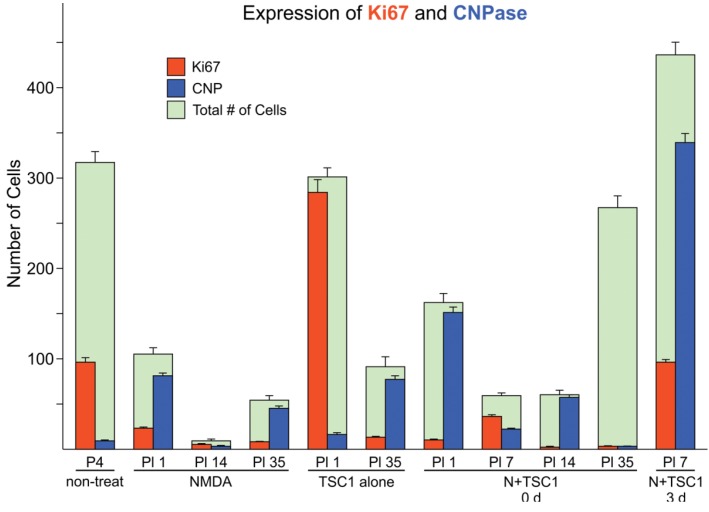
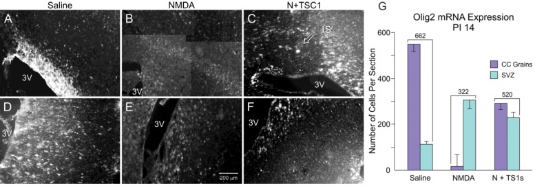
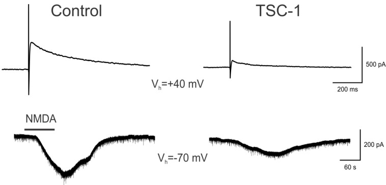
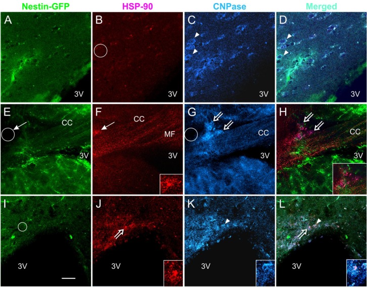
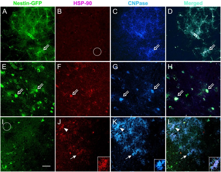
Periventricular leukomalacia (PVL) is the most frequent cause of cerebral palsy and other intellectual disabilities, and currently there is no treatment. In PVL, glutamate excitotoxicity (GME) leads to abnormal oligodendrocytes (OLs), myelin deficiency, and ventriculomegaly. We have previously identified that the combination of transferrin and insulin growth factors (TSC1) promotes endogenous OL regeneration and remyelination in the postnatal and adult rodent brain. Here, we produced a periventricular white matter lesion with a single intracerebral injection of N-methyl-d-aspartate (NMDA). Comparing lesions produced by NMDA alone and those produced by NMDA + TSC1 we found that: NMDA affected survival and reduced migration of OL progenitors (OLPs). In contrast, mice injected with NMDA + TSC1 proliferated twice as much indicating that TSC1 supported regeneration of the OLP population after the insult. Olig2-mRNA expression showed 52% OLP survival in mice receiving a NMDA injection and increased to 78% when TSC1 + NMDA were injected simultaneously and ventricular size was reduced by TSC1. Furthermore, in striatal slices TSC1 reduced the inward currents induced by NMDA in medium-sized spiny neurons, demonstrating neuroprotection. Thus, white matter loss after excitotoxicity can be partially rescued as TSC1 conferred neuroprotection to preexisting OLP and regeneration via OLP proliferation. Furthermore, we showed that early TSC1 administration maximizes neuroprotection.


Figures

Figure 1
Figure 1
The enlargement of the ventricle after excitotoxicity is decreased with a single dose of TSC1. Seven and 35 days after injection of the specific treatments para-coronal, 28 μm frozen sections were rinsed and mounted for stereological measurements. The volume of the entire ipsilateral (IL) ventricular region was selected and imaged using a 5× objective and the Cavalieri Probe (MBF) obtained in μm3. Data represent measurements of 6 adjacent coronal sections per time point from three separate experiments. The diagram shows a coronal section and the arrow points at the injection site (IS). Values are expressed as mean ± SEM of three independent experiments. p ≤ 0.05 vs. controls. All differences across treatments were significant. Abbreviations: P5 = Postnatal day five; PI = post injection day.
Figure 2
Figure 2
Cell loss in the subventricular zone (SVZ) is partially rescued in the presence of TSC1 via cell survival or cell proliferation. Double immunofluorescence: cells expressing the proliferation marker Ki67 were found in this region at early time points in the presence of TSC1. Some cells co-expressed the two markers (Ki67/CNPase). In contrast, when N-methyl-d-aspartate (NMDA) was injected alone there was a dramatic reduction of the total number of cells. Green bars represent the total number of cells (i.e., 100% or the total number of cells counted in that field. Numbers for saline and non-treated mice were very close with no significant differences. Values are expressed as mean ± SEM of the counts of 9 fields per area from three independent experiments. p < 0.05 vs. controls. All differences with respect to non-treated mouse brains as well as, across treatments were significant. P5 = Postnatal day five; PI = post injection day.
Figure 3
Figure 3
Panel (AF). More Olig 2 expressing cells survive after NMDA exposure in the presence of TSC1. Examples of expression of the transcription factor Olig2 in the corpus callosum (CC) and SVZ 14 days after injection. Animals injected with saline displayed a large number of Olig2-positive cells in the CC (A) and a reduced number in the sub-ventricular zone (SVZ, D) suggesting that the vast majority of OLP had already migrated to the CC. (B) The CC of NMDA injected mice showed fewer cells and less intensely labeled cells while some intensely labeled cells were found in the SVZ (E). (C) the CC of the mouse injected with NMDA + TSC1 showed an extensive area containing Olig2-mRNA, this particular section shows the injection site (IS) devoid of Olig2-expressing cells but surrounded by intensely labeled OL progenitors. (F) Some Olig2-expressing cells were still present in the SVZ. (C) View of the injection site at the level of the CC of a mouse co-injected with NMDA and TSC1. The intensity of the label was low and the number of cells appeared reduced in both regions when compared to saline injected mice. (G) Quantitative data of Olig2-expressing cells. In animals injected with NMDA alone, the total number of Olig2-expressing cells in the CC and SVZ was reduced when compared to the total number of cells present in mice injected with saline. In contrast, there were more Olig2-expressing cells in the SVZ, than in animals injected with saline. Interestingly, with the N + TSC1 injection the total number of Olig2-positive cells in the CC and the SVZ was partially restored to 78% and almost to an equal distribution in the SVZ and the CC, suggesting that Olig2 positive cell migration was not disrupted by NMDA in the presence of TSC1. Values are expressed as mean ± SEM of the counts of 6 fields per area from three independent experiments. p ≤ 0.05. All differences across treatments were significant.
Figure 4
Figure 4
Myelination is partially rescued from excitotoxicity by TSC1. Representative para-sagittal brain sections (28 μm thick) stained with the Spielmeyer’s method for frozen sections. These views show (A) the myelination pattern with saline treatment 40 days post-injection (PI). The extent of tissue damage, ventricle enlargement, and myelin loss in mice treated with NMDA (B) and its recovery with TSC1 (C) treatment. Moreover, NMDA treated mice showed areas where tissue was spared in the CC and CPu but there was not myelin staining. In contrast, mice treated with TSC1 showed nice myelinated fibers indicating that after the excitotoxic insult functional OLs developed and actively myelinated axons. The arrow points to a bundle of myelinated axons in the CPu of a mouse treated with NMDA + TSC1 simultaneously. In contrast, the same region of mice injected with NMDA alone did not show myelinated axons. The variability within each treatment group, consisting of 6 animals, was minimal based on low magnification examination as shown in Figure 4.
Figure 5
Figure 5
Neuroprotection of Medium-sized Spiny Neurons (MSNs). Upper traces represent responses of striatal MSNs evoked by electrical stimulation (0.2 mA, 0.1 ms duration) of cortical inputs. Recordings were obtained with patch electrodes in voltage clamp mode (holding voltage at +40 mV). Control trace is a NMDA receptor-mediated response recorded in normal ACSF solution and isolated pharmacologically by adding 2,3-dihydroxy-6-nitro-7-sulfamoyl-benzo[f]quinoxaline-2,3-dione (NBQX, 10 μM) and Bicuculline (10 μM). The trace on the right is from another cell recorded after the slice was incubated for 1 h in TSC1 solution (2 μL/mL). Lower traces show responses to bath application of NMDA (100 μM) in ACSF (left) or after incubation for 1 h in TSC-1. Notice that the NMDA responses were significantly reduced compared to control conditions. Calibration bars apply to both traces.
Figure 6
Figure 6
Acute NMDA exposure elicits the expression of HSP-90 in the CC and TSC1 neutralizes the NMDA effect. Coronal brain slices 300 μm thick were used for the acute treatment of NMDA alone or in slices pre-incubated with TSC1. After electrophysiology, slices were fixed and immunolabeled for the cell stress marker HSP-90 and the OL marker CNPase. (AD) representative views of untreated slices, neither nestin (A) nor CNPase-expressing cells (C-arrowheads) were labeled for HSP-90 (B-circle). (D) merged image. After acute NMDA, CNPase-positive OL expressed HSP-90 (EF). Moreover, cells that were not labeled for either of the two markers also expressed HSP-90 (F,G-thin arrows and circle). In slices pre-incubated with TSC1 followed by acute NMDA treatment (see methods for details) (IL) the majority of CNPase-positive cells did not express HSP-90 (K). Nestin-positive cells did not express HSP-90 in the presence of TSC1 (I-circle). Some cells co-expressed CNPase and HSP-90 (J,K and L-open arrows). Insets show higher magnification views of the cells pointed by open arrows in (J) and (L). Arrowhead points to a CNPase-positive cell (K) that co-expresses HSP-90 (L). The inset in (J) shows an example on the colocalization of both HSP90 and CNPase both, in the cell body and processes (K,L). Calibration bar in (I) corresponds to 50 μm.
Figure 7
Figure 7
Acute NMDA elicits the expression of HSP-90 in the CPu and TSC1 neutralizes the NMDA effect. Views of the CPu of 300 μm coronal slices used for the acute treatment of NMDA alone or in slices pre-incubated with TSC1. After electrophysiology, slices were fixed and immunolabeled for the cell stress marker HSP-90 and the OL marker CNPase. HSP-90 was expressed in some cells located in the CPu white matter. Non-treated slices (AD) displayed nestin-expressing cells that were negative for HSP-90 (Bempty circle). Slices directly exposed to NMDA (EH); showed colocalization of nestin and HSP-90 (E,F) and few cells co-express the three markers (open arrows). CNP-expressing cells lost processes or expression of CNPase in their processes. Slices incubated with TSC1 prior to NMDA. TSC1 appeared to maintain the tissue in a mild stage of stress with fewer nestin-expressing cells co-expressing HSP-90 (panel I) and increased number of CNPase-positive OL (K). Arrowheads show cells co-expressing HSP-90 and CNPase (K). Insets show detail of the label distribution of the cell marked with arrowheads, the merged view allows for the visualization of HSP-90 in the cell soma while CNPase is distributed in the cell body and numerous cell processes (L). K-inset shows a healthy OL that did not express HSP-90 (I-circle).

Similar articles

Cited by

References

    1. Nave K.A. Myelination and the trophic support of long axons. Nat. Rev. Neurosci. 2010;11:275–283. doi: 10.1038/nrn2797. - DOI - PubMed
    1. Blumenthal I. Periventricular leucomalacia: A review. Eur. J. Pediatr. 2004;163:435–442. doi: 10.1007/s00431-004-1477-y. - DOI - PubMed
    1. Du Plessis A.J., Volpe J.J. Perinatal brain injury in the preterm and term newborn. Curr. Opin. Neurol. 2002;15:151–157. doi: 10.1097/00019052-200204000-00005. - DOI - PubMed
    1. Goldberg M.P., Ransom B.R. New light on white matter. Stroke. 2003;34:330–332. doi: 10.1161/01.STR.0000054048.22626.B9. - DOI - PubMed
    1. Deng W. Neurobiology of injury to the developing brain. Nat. Rev. Neurol. 2010;6:328–336. doi: 10.1038/nrneurol.2010.53. - DOI - PubMed