. 2014 Mar 3;4(1):91-122.
doi: 10.3390/brainsci4010091.

Monomeric, oligomeric and polymeric proteins in huntington disease and other diseases of polyglutamine expansion

Affiliations

Monomeric, oligomeric and polymeric proteins in huntington disease and other diseases of polyglutamine expansion

Guylaine Hoffner et al. Brain Sci. .

Abstract

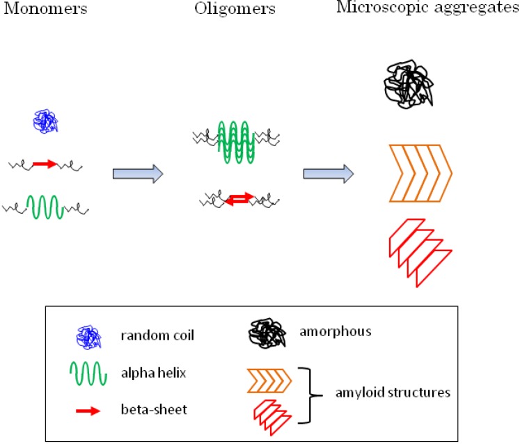
Huntington disease and other diseases of polyglutamine expansion are each caused by a different protein bearing an excessively long polyglutamine sequence and are associated with neuronal death. Although these diseases affect largely different brain regions, they all share a number of characteristics, and, therefore, are likely to possess a common mechanism. In all of the diseases, the causative protein is proteolyzed, becomes abnormally folded and accumulates in oligomers and larger aggregates. The aggregated and possibly the monomeric expanded polyglutamine are likely to play a critical role in the pathogenesis and there is increasing evidence that the secondary structure of the protein influences its toxicity. We describe here, with special attention to huntingtin, the mechanisms of polyglutamine aggregation and the modulation of aggregation by the sequences flanking the polyglutamine. We give a comprehensive picture of the characteristics of monomeric and aggregated polyglutamine, including morphology, composition, seeding ability, secondary structure, and toxicity. The structural heterogeneity of aggregated polyglutamine may explain why polyglutamine-containing aggregates could paradoxically be either toxic or neuroprotective.


Figures

Figure 1
Figure 1
Monomeric and aggregated expanded huntingtin consist of structurally heterogeneous particles. The polyQ in soluble monomeric fragments of expanded huntingtin generally adopts a random coil structure. It may also display regular structures such as α-helix or β-sheet. Oligomers may be made up of an α-helix core formed by the first seventeen amino-acids of huntingtin, with the polyQ being exposed and unstructured. Alternatively they may consist of small aggregates with buried polyQ, presumably with an antiparallel β-sheet rich structure. Microscopic aggregates (inclusions), composed of expanded huntingtin and other sequestered proteins, include amorphous aggregates and polymorphic amyloid conglomerates rich in β-sheets with antiparallel and/or parallel arrangements. Monomers, oligomers, and microscopic aggregates of expanded huntingtin, therefore, each comprise heterogeneous populations in terms of structure. They are additionally heterogeneous in size and morphology (see text).

Similar articles

Cited by

References

    1. La Spada A., Wilson E., Lubahn D., Harding A., Fischbeck K. Androgen receptor gene mutations in X-linked spinal and bulbar muscular atrophy. Nature. 1991;352:77–79. doi: 10.1038/352077a0. - DOI - PubMed
    1. The Huntington’s Disease Collaborative Research Group A novel gene containing a trinucleotide repeat that is expanded and unstable on Huntington’s disease chromosomes. Cell. 1993;72:971–983. doi: 10.1016/0092-8674(93)90585-E. - DOI - PubMed
    1. Koide R., Ikeuchi T., Onodera O., Tanaka H., Igarashi S., Endo K., Takahashi H., Kondo R., Ishikawa A., Hayashi T. Unstable expansion of cag repeat in hereditary dentatorubral-pallidoluysian atrophy (DRPLA) Nat. Genet. 1994;6:9–13. doi: 10.1038/ng0194-9. - DOI - PubMed
    1. Orr H., Chung M., Banfi S., Kwiatkowski T.J., Servadio A., Beaudet A., McCall A., Duvick L., Ranum L., Zoghbi H. Expansion of an unstable trinucleotide CAG repeat in spinocerebellar ataxia type 1. Nat. Genet. 1993;4:221–226. doi: 10.1038/ng0793-221. - DOI - PubMed
    1. Sanpei K., Takano H., Igarashi S., Sato T., Oyake M., Sasaki H., Wakisaka A., Tashiro K., Ishida Y., Ikeuchi T., et al. Identification of the spinocerebellar ataxia type 2 gene using a direct identification of repeat expansion and cloning technique, direct. Nat. Genet. 1996;14:277–284. doi: 10.1038/ng1196-277. - DOI - PubMed