Review
. 2014 Jul;141(13):2549-58.
doi: 10.1242/dev.101444.

The roles and regulation of multicellular rosette structures during morphogenesis

Affiliations
Review

The roles and regulation of multicellular rosette structures during morphogenesis

Molly J Harding et al. Development. 2014 Jul.

Abstract

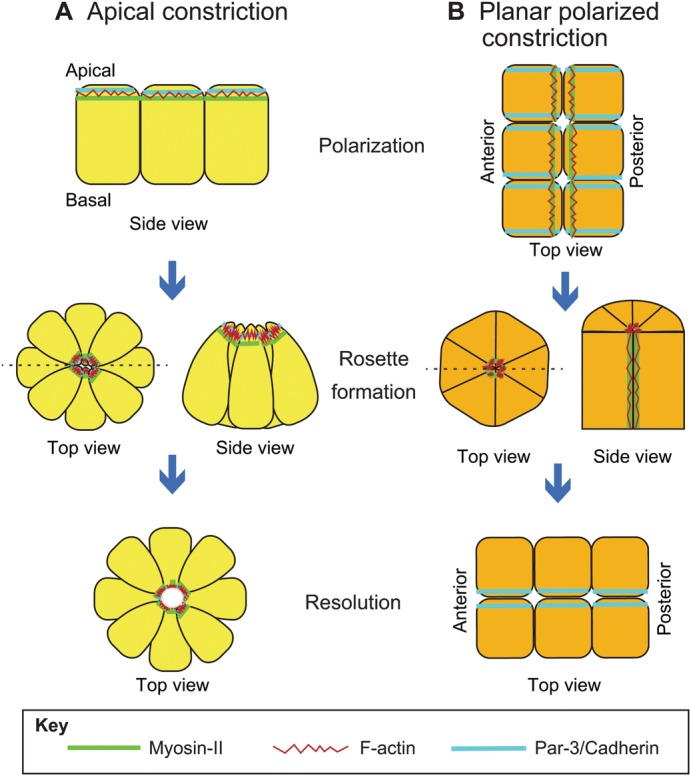
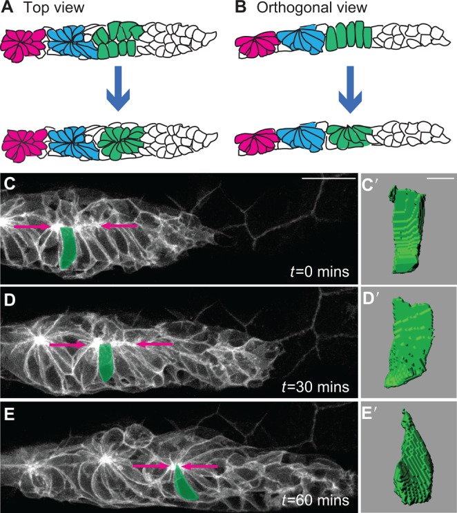
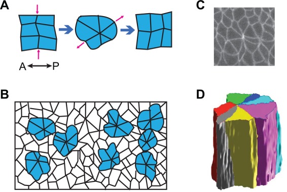
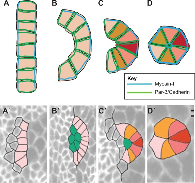
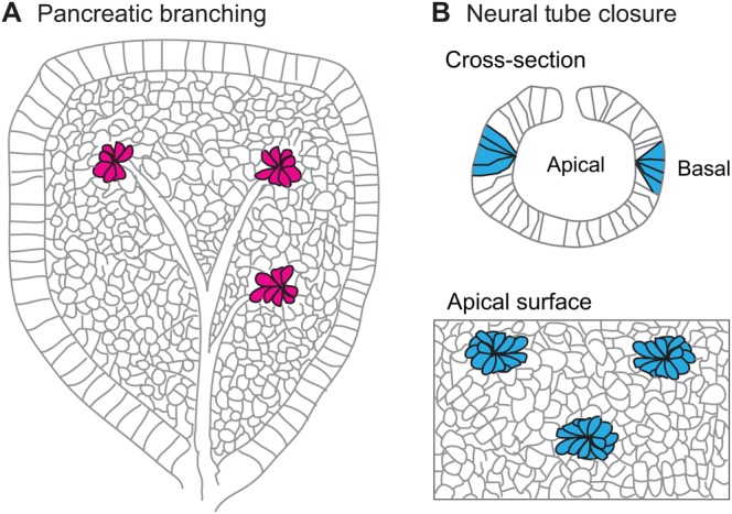
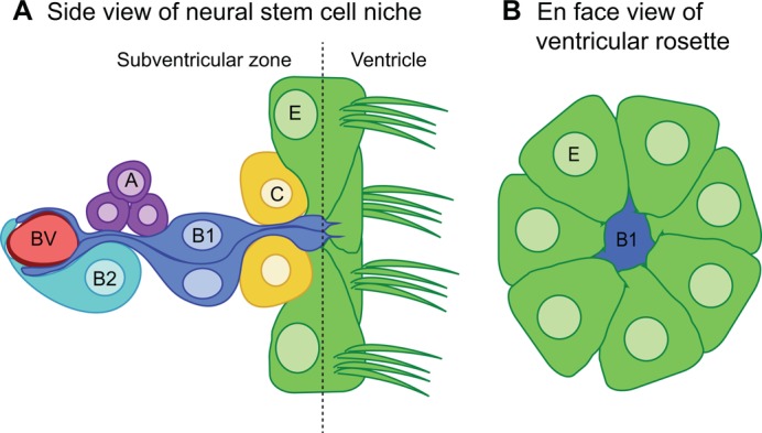
Multicellular rosettes have recently been appreciated as important cellular intermediates that are observed during the formation of diverse organ systems. These rosettes are polarized, transient epithelial structures that sometimes recapitulate the form of the adult organ. Rosette formation has been studied in various developmental contexts, such as in the zebrafish lateral line primordium, the vertebrate pancreas, the Drosophila epithelium and retina, as well as in the adult neural stem cell niche. These studies have revealed that the cytoskeletal rearrangements responsible for rosette formation appear to be conserved. By contrast, the extracellular cues that trigger these rearrangements in vivo are less well understood and are more diverse. Here, we review recent studies of the genetic regulation and cellular transitions involved in rosette formation. We discuss and compare specific models for rosette formation and highlight outstanding questions in the field.

Keywords: Drosophila epithelium; Morphogenesis; Myosin II; Rosette; Zebrafish lateral line.


Figures

Fig. 1.
Fig. 1.
Mechanisms of rosette formation. (A) Rosette formation via apical constriction. Prior to apical constriction, cytoskeletal molecules, including F-actin and myosin-II, Par-3 and N-cadherin, are in the apical domains of columnar cells. Apical constriction leads to the formation of a rosette around an acto-myosin-rich center. Note that only apical, but not basal, domains of rosette cells are constricted. Following rosette formation, rosette centers may open to form a central lumen. (B) During rosette formation via planar polarized constriction, cytoskeletal molecules are distributed in a planar polarized fashion throughout the developing tissue, prior to rosette formation. Actin and myosin-II are localized to the AP axis, and Par-3 (Baz) and cadherin are localized to perpendicular membranes. This distribution allows the formation of a rosette with a myosin-II-rich center. Note that cells constrict along their lateral edges. Rosettes formed in this manner often resolve along the axis perpendicular to that of the initial cellular arrangement. Dashed lines indicate the plane of the cross-sections shown.
Fig. 2.
Fig. 2.
Rosette formation in the zebrafish posterior lateral line. (A,B) In the zebrafish posterior lateral line (pLL), rosettes form in the leading (caudal) part of the pLL primordium, where columnar cells constrict apically to form a new rosette. Note that the apical ends of teardrop-shaped cells are oriented towards the rosette center. (C-E) Live imaging of rosette formation in a Tg(claudinB:lynEGFP) zebrafish embryo (Haas and Gilmour, 2006; modified with permission from Harding and Nechiporuk, 2012). Over the course of 1 hour, cells apically constrict (arrows) to form a rosette. (C′-E′) Three-dimensional reconstructions of the pseudocolored cell in C-E demonstrate progressive apical constriction of rosette-forming cells over time. Scale bars: 20 μm in C-E; 4 μm in C′-E′.
Fig. 3.
Fig. 3.
Rosette formation during Drosophila epithelial elongation. (A) Rosettes form and resolve along perpendicular axes to drive cellular rearrangement and epithelial elongation. A, anterior; P, posterior. (B) Schematized apical view of the epithelium. Note that many cells in the epithelium participate in rosette formation (see Blankenship et al., 2006). (C) A rosette in a wild-type Drosophila embryo expressing Resille:GFP, a membrane marker. (D) Three-dimensional reconstruction of an epithelial rosette. Note that in this rosette type, cells are joined along their lateral lengths, in contrast to apically constricted rosettes.
Fig. 4.
Fig. 4.
Rosette formation during Drosophila ommatidial development. (A) Lines of up to ten cells form prior to rosette formation, with complementary localization of myosin-II and Par-3/cadherin. (B-D) Cells transition into arcs (B,C), which subsequently form the five-cell rosette (D), which contains a myosin-II-rich rosette center. During rosette formation, ommatidial cell fate also begins to be specified; the different colors represent distinct cell fates. (A′-D′) Formation of lines (A′), arcs (B′,C′) and rosettes (D′) in the eye disc (modified with permission from Robertson et al., 2012).
Fig. 5.
Fig. 5.
Rosette formation during pancreatic branching morphogenesis and neural tube closure. (A) Rosettes in the developing pancreas. The rosettes formed subsequently open up to form microlumens. Modified with permission from Villasenor et al. (2010). (B) Rosettes in the developing neural tube form via apical constriction. Multiple rosettes are visible simultaneously in the apical surface of the neural tube.
Fig. 6.
Fig. 6.
Rosette structures in the adult neural stem cell niche. (A) The ventricular-subventricular (V-SVZ) adult neural stem cell niche. The niche is composed of ependymal (E) cells on the ventricular wall and astrocyte-like stem cells (B1 and B2) that, in the case of type-B1 cells, extend from the ventricular space to the subventricular space and make contact with blood vessels (BV) of the central nervous system. Type-B1 and -B2 cells give rise to rapidly proliferating progenitor cells (C and A) that will form new neurons and glial cells. (B) En face view of the lateral ventricular wall showing the rosette arrangement of ependymal cells surrounding the apical tips of type-B1 cells. See Fuentealbe et al. (2012).

Similar articles

Cited by

References

    1. Afonso C., Henrique D. (2006). PAR3 acts as a molecular organizer to define the apical domain of chick neuroepithelial cells. J. Cell Sci. 119, 4293-4304 10.1242/jcs.03170 - DOI - PubMed
    1. Baonza A., Casci T., Freeman M. (2001). A primary role for the epidermal growth factor receptor in ommatidial spacing in the Drosophila eye. Curr. Biol. 11, 396-404 10.1016/S0960-9822(01)00125-7 - DOI - PubMed
    1. Bertet C., Sulak L., Lecuit T. (2004). Myosin-dependent junction remodelling controls planar cell intercalation and axis elongation. Nature 429, 667-671 10.1038/nature02590 - DOI - PubMed
    1. Blankenship J. T., Backovic S. T., Sanny J. S. P., Weitz O., Zallen J. A. (2006). Multicellular rosette formation links planar cell polarity to tissue morphogenesis. Dev. Cell 11, 459-470 10.1016/j.devcel.2006.09.007 - DOI - PubMed
    1. Brasher B. B., Van Etten R. A. (2000). c-Abl has high intrinsic tyrosine kinase activity that is stimulated by mutation of the Src homology 3 domain and by autophosphorylation at two distinct regulatory tyrosines. J. Biol. Chem. 275, 35631-35637 10.1074/jbc.M005401200 - DOI - PubMed

Publication types

LinkOut - more resources