Review
. 2014 Jul;141(13):2559-67.
doi: 10.1242/dev.104588.

Heterogeneity and plasticity of epidermal stem cells

Affiliations
Review

Heterogeneity and plasticity of epidermal stem cells

Troels Schepeler et al. Development. 2014 Jul.

Abstract

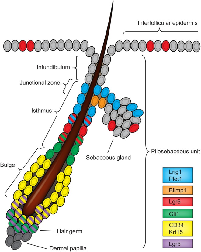
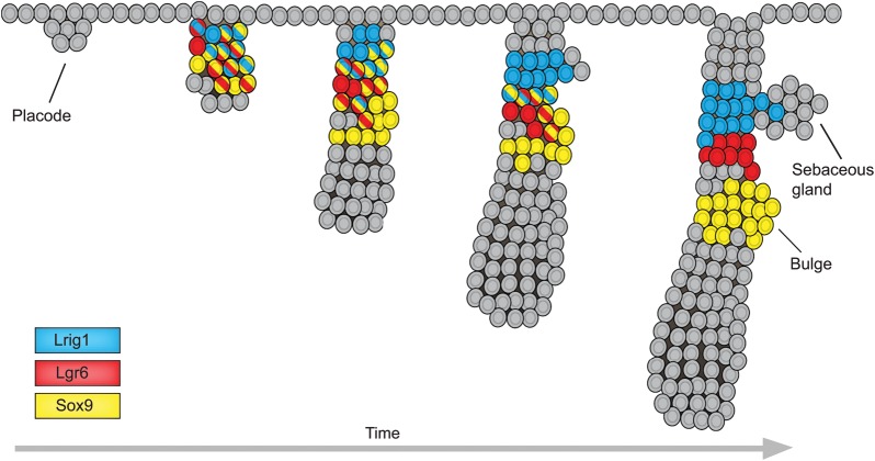
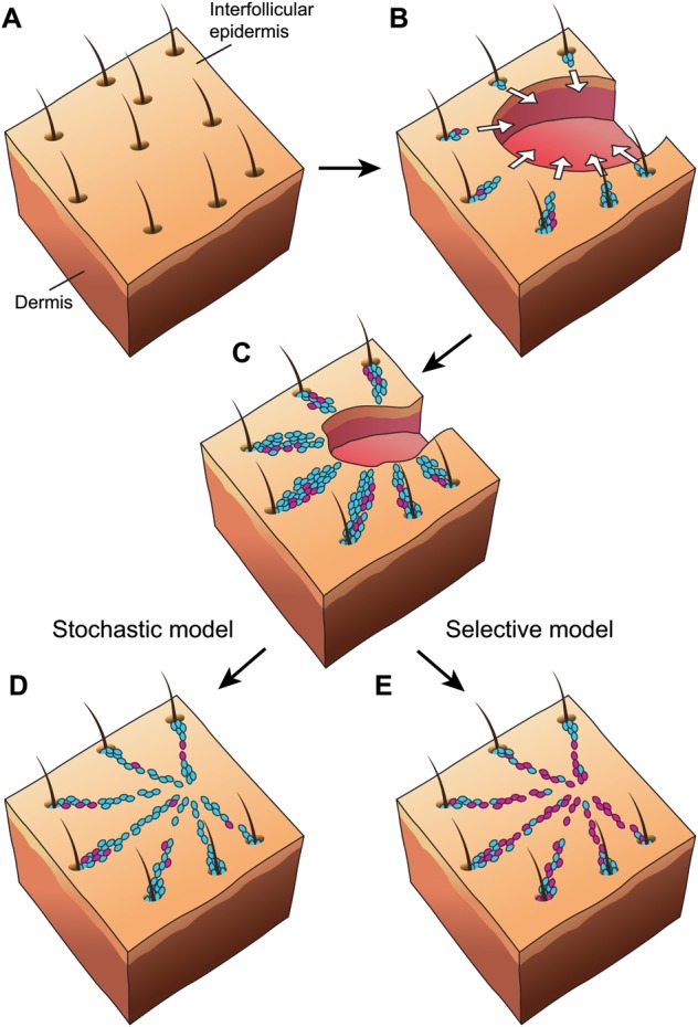
The epidermis is an integral part of our largest organ, the skin, and protects us against the hostile environment. It is a highly dynamic tissue that, during normal steady-state conditions, undergoes constant turnover. Multiple stem cell populations residing in autonomously maintained compartments facilitate this task. In this Review, we discuss stem cell behaviour during normal tissue homeostasis, regeneration and disease within the pilosebaceous unit, an integral structure of the epidermis that is responsible for hair growth and lubrication of the epithelium. We provide an up-to-date view of the pilosebaceous unit, encompassing the heterogeneity and plasticity of multiple discrete stem cell populations that are strongly influenced by external cues to maintain their identity and function.

Keywords: Epidermis; Regeneration; Stem cells; Tissue homeostasis.


Figures

Fig. 1.
Fig. 1.
Hair follicle stem cell compartments. Different stem cell populations are shown in the mouse resting (telogen) adult hair follicle. Each stem cell compartment is defined by distinct protein expression and gene promoter activity (see key); cells with multiple colours express multiple markers.
Fig. 2.
Fig. 2.
Emergence of distinct stem cell populations during morphogenesis of the pilosebaceous unit. During development, pilosebaceous formation is initiated from an early epidermal structure (the placode) that develops into a fully formed pilosebaceous unit (PSU) through a series of steps involving complex interactions with existing dermal cells. Initially, various stem cell markers are co-expressed within the same region of the developing PSU, but at later stages marker expression is associated with segregation of cells into distinct domains. Cells with multiple colours express multiple markers.
Fig. 3.
Fig. 3.
Mobilisation of stem cell progeny from the PSU following wounding. Upon full-thickness wounding of intact skin (A), stem cell progeny from multiple PSUs along the wound edge migrate toward the centre of the wound area (white arrows, B) to participate in the regenerative response within the interfollicular epidermis. Two lineage-marked populations represented by blue cells and violet cells are depicted. Re-epithelialisation of the wound area occurs with a larger fraction of blue cell progeny present than violet cell progeny, which can result from the differential timing of stem cell activation or different labelling efficiencies, as shown in C. According to a stochastic model (D), the long-term contribution of stem cell progeny in the wound area is proportional to their initial representation in the wound epidermis. Since there is no difference in the fitness between the blue and violet cells, blue cells outnumber violet cells because of their initial numerical advantage. By contrast, a selective model (E) assumes that intrinsic fitness differences between stem cell progeny do exist. Here, violet cells have an advantage compared with blue cells, so despite being under-represented they eventually dominate the wound area.

References

    1. Agrawal N., Frederick M. J., Pickering C. R., Bettegowda C., Chang K., Li R. J., Fakhry C., Xie T.-X., Zhang J., Wang J., et al. (2011). Exome sequencing of head and neck squamous cell carcinoma reveals inactivating mutations in NOTCH1. Science 333, 1154-1157 10.1126/science.1206923 - DOI - PMC - PubMed
    1. Ahmed M. I., Mardaryev A. N., Lewis C. J., Sharov A. A., Botchkareva N. V. (2011). MicroRNA-21 is an important downstream component of BMP signalling in epidermal keratinocytes. J. Cell Sci. 124, 3399–3404 10.1242/jcs.086710 - DOI - PMC - PubMed
    1. Alcolea M. P., Jones P. H. (2014). Lineage analysis of epidermal stem cells. Cold Spring Harb. Perspect. Med. 4, a015206 10.1101/cshperspect.a015206 - DOI - PMC - PubMed
    1. Alonso L., Fuchs E. (2006). The hair cycle. J. Cell Sci. 119, 391–393 10.1242/jcs02793 - DOI - PubMed
    1. Arwert E. N., Hoste E., Watt F. M. (2012). Epithelial stem cells, wound healing and cancer. Nat. Rev. Cancer 12, 170-180 10.1038/nrc3217 - DOI - PubMed

Publication types

MeSH terms