

Immunology of Taenia solium taeniasis and human cysticercosis
- PMID: 24962350
- PMCID: PMC5761726
- DOI: 10.1111/pim.12126
Immunology of Taenia solium taeniasis and human cysticercosis
Abstract
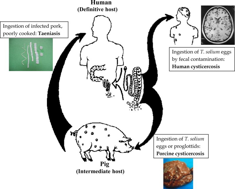
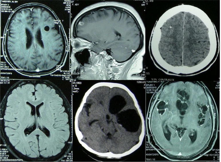
The life cycle of Taenia solium, the pork tapeworm, is continuously closed in many rural settings in developing countries when free roaming pigs ingest human stools containing T. solium eggs and develop cysticercosis, and humans ingest pork infected with cystic larvae and develop intestinal taeniasis, or may also accidentally acquire cysticercosis by faecal-oral contamination. Cysticercosis of the human nervous system, neurocysticercosis, is a major cause of seizures and other neurological morbidity in most of the world. The dynamics of exposure, infection and disease as well as the location of parasites result in a complex interaction which involves immune evasion mechanisms and involutive or progressive disease along time. Moreover, existing data are limited by the relative lack of animal models. This manuscript revises the available information on the immunology of human taeniasis and cysticercosis.
Keywords: Peru; Taenia solium; cysticercosis; neurocysticercosis; parasitic infections; seizures.
© 2014 John Wiley & Sons Ltd.
Figures
Similar articles
-
Taenia solium taeniasis/cysticercosis: From parasite biology and immunology to diagnosis and control.Adv Parasitol. 2021;112:133-217. doi: 10.1016/bs.apar.2021.03.003. Epub 2021 Apr 8. Adv Parasitol. 2021. PMID: 34024358
-
Zoonotic Taenia infections with focus on cysticercosis due to Taenia solium in swine and humans.Res Vet Sci. 2021 Jan;134:69-77. doi: 10.1016/j.rvsc.2020.11.015. Epub 2020 Nov 22. Res Vet Sci. 2021. PMID: 33321377 Review.
-
Insights into the diagnosis, vaccines, and control of Taenia solium, a zoonotic, neglected parasite.Parasit Vectors. 2023 Oct 24;16(1):380. doi: 10.1186/s13071-023-05989-6. Parasit Vectors. 2023. PMID: 37876008 Free PMC article. Review.
-
Control of the taeniosis/cysticercosis complex: future developments.Vet Parasitol. 2006 Jul 31;139(4):283-92. doi: 10.1016/j.vetpar.2006.04.019. Epub 2006 May 24. Vet Parasitol. 2006. PMID: 16730125 Review.
-
Seroepidemiological evidence for Taenia solium taeniasis/cysticercosis in three Venezuelan rural communities.J Helminthol. 2020 Aug 11;94:e179. doi: 10.1017/S0022149X20000619. J Helminthol. 2020. PMID: 32778183
Cited by
-
The effectiveness of anti-inflammatory and anti-seizure medication for individuals with single enhancing lesion neurocysticercosis: A meta-analysis and expert group-based consensus recommendations.PLoS Negl Trop Dis. 2021 Mar 31;15(3):e0009193. doi: 10.1371/journal.pntd.0009193. eCollection 2021 Mar. PLoS Negl Trop Dis. 2021. PMID: 33788843 Free PMC article.
-
Helminthic dehydrogenase drives PGE2 and IL-10 production in monocytes to potentiate Treg induction.EMBO Rep. 2022 May 4;23(5):e54096. doi: 10.15252/embr.202154096. Epub 2022 Mar 31. EMBO Rep. 2022. PMID: 35357743 Free PMC article.
-
Foodborne Parasitic Diseases in the Neotropics - A Review.Helminthologia. 2021 Jun 8;58(2):119-133. doi: 10.2478/helm-2021-0022. eCollection 2021 Jun. Helminthologia. 2021. PMID: 34248373 Free PMC article.
-
Urinary neopterin reflects immunological variation associated with age, helminth parasitism, and the microbiome in a wild primate.Sci Rep. 2022 Dec 9;12(1):21307. doi: 10.1038/s41598-022-25298-9. Sci Rep. 2022. PMID: 36494454 Free PMC article.
-
Prevalence and risk factors for Taenia solium cysticercosis in school-aged children: A school based study in western Sichuan, People's Republic of China.PLoS Negl Trop Dis. 2018 May 8;12(5):e0006465. doi: 10.1371/journal.pntd.0006465. eCollection 2018 May. PLoS Negl Trop Dis. 2018. PMID: 29738570 Free PMC article.
References
-
- Garcia HH, Del Brutto OH. Neurocysticercosis: updated concepts about an old disease. Lancet Neurol. 2005;4(10):653–61. - PubMed
-
- Flisser A. Taeniasis and cysticercosis due to Taenia solium. Prog Clin Parasitol. 1994;4:77–116. - PubMed
-
- Schantz PM. Taenia solium cysticercosis: an overview of global distribution and transmission. In: Singh G, Prabhakar S, editors. Taenia solium cysticercosis From basic to clinical science. Oxon, UK: CABI Publishing; 2002.
Publication types
MeSH terms
Grants and funding
LinkOut - more resources
Full Text Sources
Other Literature Sources
