. 2014 Aug 15;289(33):23056-23064.
doi: 10.1074/jbc.M114.574210. Epub 2014 Jun 24.

CRL4Cdt2 E3 ubiquitin ligase and proliferating cell nuclear antigen (PCNA) cooperate to degrade thymine DNA glycosylase in S phase

Affiliations

CRL4Cdt2 E3 ubiquitin ligase and proliferating cell nuclear antigen (PCNA) cooperate to degrade thymine DNA glycosylase in S phase

Etsuko Shibata et al. J Biol Chem. .

Abstract

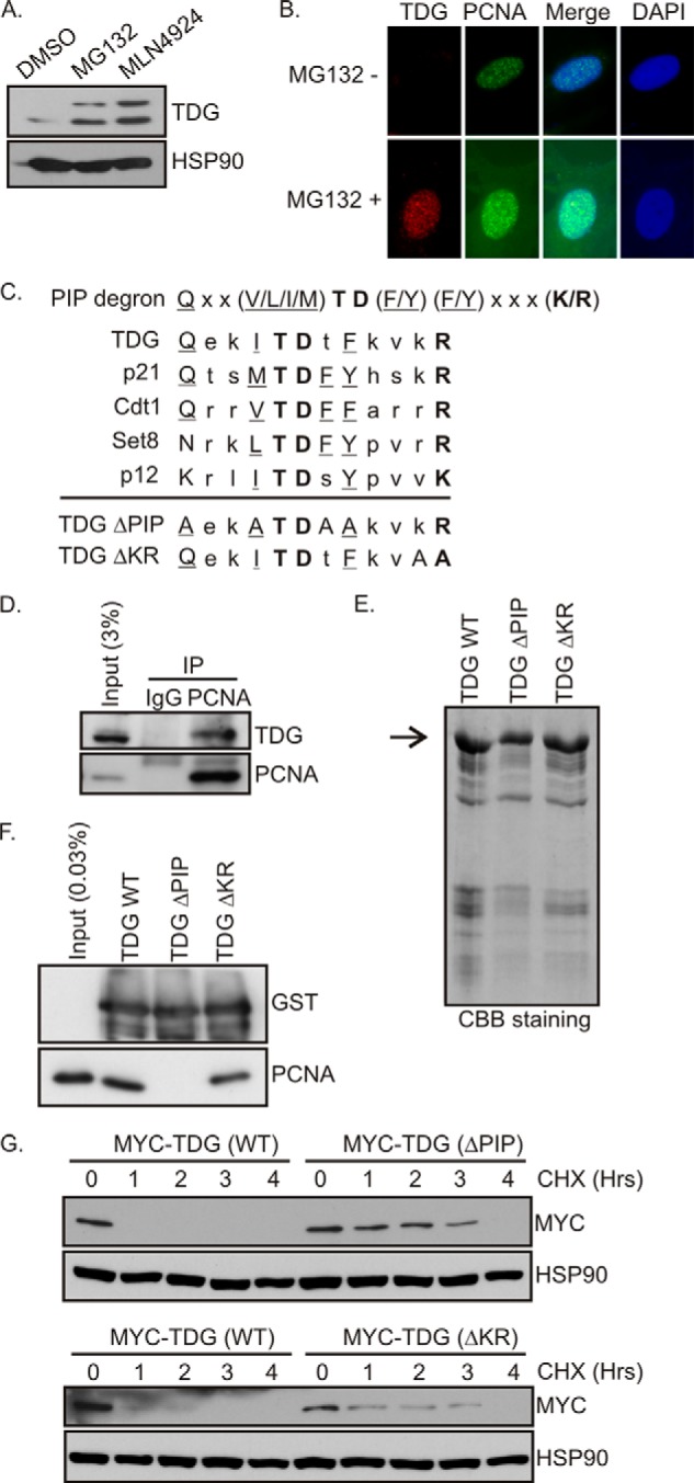
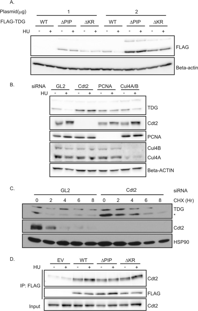
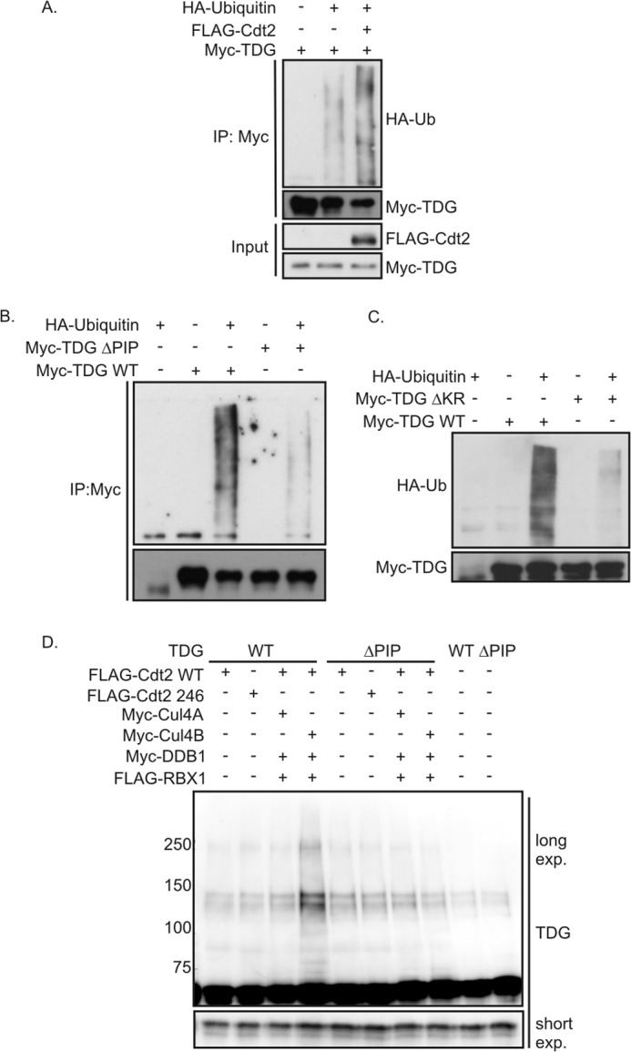
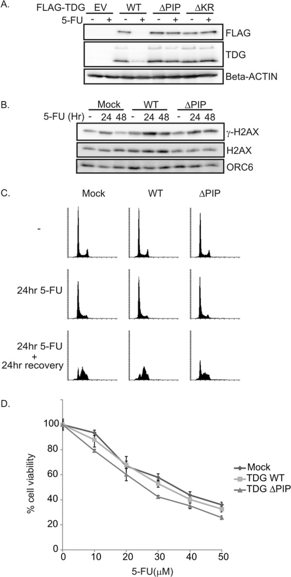
Thymine DNA glycosylase (TDG) is an essential enzyme playing multiple roles in base excision repair, transcription regulation, and DNA demethylation. TDG mediates the cytotoxicity of the anti-cancer chemotherapeutic drug 5-fluorouracil (5-FU) by prolonging S phase, generating DNA strand breaks, and inducing DNA damage signaling. During S phase of the cell cycle, TDG is degraded via the proteasomal pathway. Here we show that CRL4(Cdt2) E3 ubiquitin ligase promotes ubiquitination and proteasomal degradation of TDG in S phase in a reaction that is dependent on the interaction of TDG with proliferating cell nuclear antigen (PCNA). siRNA-mediated depletion of PCNA or components of CRL4(Cdt2), specifically cullin4A/B or substrate adaptor Cdt2, stabilizes TDG in human cells. Mutations in the PCNA-interacting peptide (PIP) motif of TDG that disrupt the interaction of TDG with PCNA or change critical basic residues essential for the action of the PIP degron prevent the ubiquitination and degradation of TDG. Thus physical interaction of TDG with PCNA through the PIP degron is required for targeting TDG to the CRL4(Cdt2) E3 ubiquitin ligase complex. Compared with forced expression of wild type TDG, CRL4(Cdt2)- resistant TDG (ΔPIP) slows cell proliferation and slightly increases the toxicity of 5-FU. Thus, CRL4(Cdt2)-dependent degradation of TDG occurs in S phase because of the requirement for TDG to interact with chromatin-loaded PCNA, and this degradation is important for preventing toxicity from excess TDG.

Keywords: DNA Damage Response; E3 Ubiquitin Ligase; Proliferating Cell Nuclear Antigen (PCNA); Protein Degradation; Thymine-DNA Glycosylase (TDG); Ubiquitin.


Figures

FIGURE 1.
FIGURE 1.
TDG interacts with PCNA. A, MG132 and MLN4924 stabilize TDG. HeLa cells (asynchronous) were treated with DMSO, MG132 (20 μm), or MLN4924 (5 μm) for 6 h. The cell lysates were probed with the indicated antibodies. B, immunofluorescence of TDG and PCNA. TDG staining is absent in the cells with S phase-specific PCNA foci unless cells are treated with MG132. C, PIP degron of CRL4 Cdt2 substrates. Conserved residues in the canonical PIP box are underlined. Conserved residues in the PIP degron are in boldface type. The mutations made in TDG(ΔPIP) and TDG(ΔKR) are shown. D, TDG interacts with PCNA. Cell extracts from HeLa cells treated with 40 μm MG132 with or without 2 mm HU were immunoprecipitated (IP) with anti-PCNA or control IgG antibodies, and the precipitates were immunoblotted with anti-TDG and anti-PCNA antibodies. E, purification of recombinant proteins. Coomassie Brilliant Blue staining of gel showing the purification of GST-TDG(WT), GST-TDG(ΔPIP), and GST-TDG(ΔKR). F, in vitro GST pull-down assay. Purified PCNA was incubated either with GST-TDG(WT) or its mutants immobilized on glutathione beads. After washing, proteins bound to beads were resolved on 10% SDS gel, and the blot was probed with anti-PCNA and GST antibodies. G, mutations in the PIP degron increase half-life of TDG. 293T cells were transfected with Myc-TDG(WT), Myc-TDG(ΔPIP), or Myc-TDG(ΔKR). After 48 h of transfection, cells were treated with cycloheximide (50 μg/ml) and harvested at different time points as indicated. The blots were probed with anti-Myc antibodies.
FIGURE 2.
FIGURE 2.
CRL4Cdt2 is required for the degradation of TDG. A, TDG mutants were not degraded after HU treatment. 293T cells in 6-well plates were transiently transfected with 1 or 2 μg of TDG(WT)-, TDG(ΔPIP)-, or TDG(ΔKR)-expressing plasmids for 24 h, followed by treatment with 1.5 mm HU for 24 h. Western blot analyses were performed using the indicated antibodies. B, HeLa cells were treated with the indicated siRNA. siRNA against luciferase (GL2) was used as a negative control. After 48 h, 1.5 mm HU was added for 24 h. Cell lysates were subjected to immunoblotting with anti-TDG and anti-β-actin antibodies. C, knockdown of Cdt2 increased the half-life of TDG. HeLa cells were treated with GL2 siRNA or siRNA against Cdt2 for 60 h, followed by protein synthesis inhibitor cycloheximide treatment (100 μg/ml) for the indicated times. The whole cell lysate was probed with antibodies against indicated proteins. *, lower band is TDG. D, TDG(WT) and mutants co-immunoprecipitate with Cdt2. Nuclear fraction of 293T cells transiently expressing the indicated forms of FLAG-TDG were immunoprecipitated (IP) with anti-FLAG. Association was analyzed by Western blot using anti-Cdt2 and anti-FLAG antibodies. EV, empty vector.
FIGURE 3.
FIGURE 3.
CRL4Cdt2 ubiquitinates TDG. A, in vivo ubiquitination assay of TDG. Myc-TDG was transfected into 293T cells with or without FLAG-Cdt2 and HA-ubiquitin. Cells were lysed under denaturing conditions and subjected to immunoprecipitation (IP) with anti-Myc and processed for immune blotting with the indicated antibodies. B and C, in vivo ubiquitination assay. Myc TDG WT or mutants were transfected into 293T cells with or without HA-ubiquitin. Cells were lysed under denaturing conditions and subjected to immune precipitation with anti-Myc and processed for immune blotting with the indicated antibodies. The exposure time is longer than in A to better see the polyubiquitination carried out by endogenous Cdt2. D, in vitro ubiquitination assay. Myc TDG was immunopurified from 293T cells to use as substrate. CRL4Cdt2 complex created by transiently overexpressing FLAG-Cdt2, FLAG-RBX1, Myc-DDB1 with Myc-Cul4A, or Myc-Cul4B was immunopurified with anti-FLAG from 293T cells and used as E3 ligase. Anti-TDG Western blot was performed on the reaction products to detect high molecular weight forms of TDG.
FIGURE 4.
FIGURE 4.
TDG overexpression decreases cell proliferation, increases S phase population, and increases DNA breaks. A, Western blot analysis of doxycycline-induced TDG expression in stably transfected HeLa cell lines. B, top, doxycycline-induced TDG overexpression reduces cell growth. Cell growth was assessed by an MTT assay. Bottom, TDG(ΔKR) is not as toxic as TDG(ΔPIP). Cells grown in the absence of doxycycline still express some TDG from leaky expression (Western blot). C, TDG-overexpressing cells accumulate in S phase. Cells were treated with 50 ng/ml of doxycycline for 3 days to induce TDG(WT), TDG(ΔPIP), or TDG(ΔKR). Cell cycle profile was analyzed by flow cytometry of propidium iodide-stained cells. Left, changes in the percentage of S phase cells induced by doxycycline, relative to cells not treated with doxycycline, are plotted (mean ± S.D. (error bars) of >3 measurements). Right, percentage of cells in each phase of the cell cycle in three independent experiments. D, increase of TDG increases DNA breaks. Cells (transfected with TDG(WT) or mock-transfected) were treated with 100 ng/ml doxycycline for 3 days. DNA breaks were analyzed by an alkaline comet assay. A box and whiskers plot (median and interquartile range) shows the yield of strand breaks as evaluated from the tail moment (n > 100 cells).
FIGURE 5.
FIGURE 5.
TDG(ΔPIP)-expressing cells are slightly sensitive to 5-FU. A, TDG mutants were not degraded upon DNA damage with 5-FU treatment. HeLa cell lines expressing TDG(WT) or mutants were exposed to 5-FU for 20 h. Western blot analyses were carried out using the indicated antibodies. B, delayed repair of DNA damage induced by 5-FU in HeLa cells expressing TDG(WT) or TDG (ΔPIP). Cells were exposed to 20 μm 5-FU for 24 h followed by a 24-h recovery period. P-H2AX (Ser-139) and H2AX levels were analyzed by Western blot. ORC6 was used as a loading control. C, TDG(ΔPIP) expression slows down resumption of S phase after G1/S arrest induced by 5-FU treatment. HeLa cells expressing TDG(WT) or TDG(ΔPIP) were exposed to 20 μm 5-FU for 24 h, followed by a 24-h recovery period. Cell cycle profile was analyzed by flow cytometry of propidium iodide-stained cells. D, HeLa cells expressing TDG(WT) or TDG(ΔPIP) were cultured with the indicated concentrations of 5-FU for 24 h, and cell viability was assessed on day 5 with the MTT assay. The relative cell viability was expressed as a percentage relative to the untreated cells. Error bars, S.D.

Similar articles

Cited by

References

    1. Hendrich B., Hardeland U., Ng H. H., Jiricny J., Bird A. (1999) The thymine glycosylase MBD4 can bind to the product of deamination at methylated CpG sites. Nature 401, 301–304 - PubMed
    1. Haushalter K. A., Todd Stukenberg M. W., Kirschner M. W., Verdine G. L. (1999) Identification of a new uracil-DNA glycosylase family by expression cloning using synthetic inhibitors. Curr. Biol. 9, 174–185 - PubMed
    1. Hardeland U., Bentele M., Lettieri T., Steinacher R., Jiricny J., Schar P. (2001) Thymine DNA glycosylase. Prog. Nucleic Acids Res. Mol. Biol. 68, 235–253 - PubMed
    1. Slupphaug G., Eftedal I., Kavli B., Bharati S., Helle N. M., Haug T., Levine D. W., Krokan H. E. (1995) Properties of a recombinant human uracil-DNA glycosylase from the UNG gene and evidence that UNG encodes the major uracil-DNA glycosylase. Biochemistry 34, 128–138 - PubMed
    1. Hardeland U., Steinacher R., Jiricny J., Schär P. (2002) Modification of the human thymine-DNA glycosylase by ubiquitin-like proteins facilitates enzymatic turnover. EMBO J. 21, 1456–1464 - PMC - PubMed

Publication types

MeSH terms

LinkOut - more resources