. 2014 Aug 8;289(32):22048-62.
doi: 10.1074/jbc.M114.568790. Epub 2014 Jun 24.

Sirtuin-1 (SIRT1) is required for promoting chondrogenic differentiation of mesenchymal stem cells

Affiliations

Sirtuin-1 (SIRT1) is required for promoting chondrogenic differentiation of mesenchymal stem cells

Constanze Buhrmann et al. J Biol Chem. .

Abstract

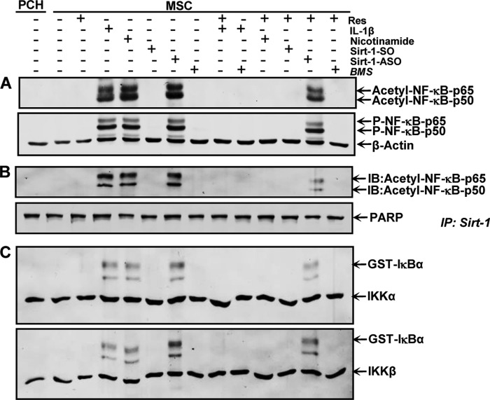
Sirtuin-1 (SIRT1), NAD(+)-dependent deacetylase, has been linked to anabolic effects in cartilage, although the mechanisms of SIRT1 signaling during differentiation of mesenchymal stem cells (MSCs) to chondrocytes are poorly understood. Therefore, we investigated the role of SIRT1-mediated signaling during chondrogenic differentiation of MSCs in vitro. High density and alginate cultures of MSCs were treated with chondrogenic induction medium with/without the SIRT1 inhibitor nicotinamide, antisense oligonucleotides against SIRT1 (SIRT1-ASO), IL-1β, and/or resveratrol. Transient transfection of MSCs with SIRT1-antisense oligonucleotides, nicotinamide, and IL-1β inhibited chondrogenesis-induced down-regulation of cartilage-specific proteins, cartilage-specific transcription factor Sox9, and enhanced NF-κB-regulated gene products involved in the inflammatory and degradative processes in cartilage (MMP-9, COX-2, and caspase-3), and NF-κB phosphorylation, acetylation, and activation of IκBα kinase. In contrast, the SIRT1 activator resveratrol or BMS-345541 (inhibitor of IKK) inhibited IL-1β- and NAM-induced suppression of cartilage-specific proteins, Sox9, and up-regulation of NF-κB-regulated gene products. Moreover, SIRT1 was found to interact directly with NF-κB and resveratrol-suppressed IL-1β and NAM but not SIRT1-ASO-induced NF-κB phosphorylation, acetylation, and activation of IκBα kinase. Knockdown of SIRT1 by mRNA abolished the inhibitory effects of resveratrol on inflammatory and apoptotic signaling and Sox9 expression, suggesting the essential role of this enzyme. Finally, the modulatory effects of resveratrol were found to be mediated at least in part by the association between SIRT1 and Sox9. These results indicate for the first time that SIRT1 supports chondrogenic development of MSCs at least in part through inhibition/deacetylation of NF-κB and activation of Sox9.

Keywords: Chondrogenesis; Inflammation; Mesenchymal Stem Cells (MSCs); NF-κB; Resveratrol; Rheumatoid Arthritis; SIRT1-ASO; SOX9; Sirtuin 1 (SIRT1).


Figures

FIGURE 1.
FIGURE 1.
Characterization of MSCs. A, in monolayer culture the human MSCs (panel a) showed a polymorphic, fibroblast-like morphology. The MSCs expressed positive stem cell-specific markers CD90+ (panel b) and CD105+ (panel c) and were negative for the hematopoietic stem cell markers CD45 (panel d) and CD34 (panel e). Magnification: ×10 (panel a), ×40 (panels b–e), and bar, 1 μm. B, to quantify the stem cell marker-positive cells, the labeled cultures were examined by counting 300 cells from 10 microscopic fields. The examination was performed in triplicate, and the results are provided as the mean values with S.D. from three independent experiments. Values were compared with the control, and statistically significant values with p < 0.05 are designated by a star, and p < 0.01 is designated by two stars. Co., without primary antibody, followed by incubation with rhodamine-coupled secondary antibodies and counterstaining with DAPI to visualize the cell nuclei.
FIGURE 2.
FIGURE 2.
Effects of SIRT1-ASO on chondrogenic differentiation of MCS in vitro. A, primary human chondrocytes (PCH) in high density were left untreated (panel a). MSCs were either cultured only with chondrogenic induction medium (panel b) or with chondrogenic induction medium and simultaneously transfected with different concentrations (0.1, 0.5, 1, 5 μm) of SIRT1-SO (panels c–f) or SIRT1-ASO (panels g–j) in the presence of Lipofectin (10 μl/ml). Ultrastructural morphology of high density cultures was evaluated after 14 days by transmission electron microscope. Control cultures of chondrocytes or MSCs or treatment of MSCs with SIRT1-SO showed well developed chondrocytes (Ch) embedded in a well developed ECM and the formation of cartilage nodules (panels a–f). Treatment with SIRT1-ASO resulted in matrix breakdown and cell lysis and apoptosis (arrows) (panels g–j). Micrographs shown are representative of three individual experiments. Magnification: ×5000, bar, 1 μm. B, to quantify degenerative changes, high density cultures of A were examined for apoptosis by counting 100 cells from 25 microscopic fields. The examination was performed in triplicate, and the results are provided as the mean values with S.D. from three independent experiments. Values were compared with the control and statistically significant values with p < 0.05 were designated by a star and p < 0.01 were designated by a two stars.
FIGURE 3.
FIGURE 3.
Effects of SIRT1-ASO on SIRT1 expression, ECM protein production, and cartilage-specific transcription factor Sox9 during chondrogenesis of MSC in vitro. Primary human chondrocytes (PCH) in high density were left untreated. MSCs in high density were either cultured only with chondrogenic induction medium or with chondrogenic induction medium and transfected with different concentrations of SIRT1-SO or SIRT1-ASO (0.1, 0.5, 1, and 5 μm) in the presence of Lipofectin (10 μl/ml) for 14 days. Whole cell lysates were prepared and analyzed by Western blotting with antibodies against SIRT1 (A), collagen type II (B), cartilage-specific proteoglycans (C), and cartilage-specific transcription factor Sox9 (D). Densitometric evaluation of protein expression as revealed by Western blot analysis was performed in triplicate. Bars represent the mean values (± S.D.) for collagen type II of the 216-, 137-, and 110-kDa bands and for cartilage-specific proteoglycans of the 140-, 127-, and 33-kDa bands. SIRT1 control peptide (Co. Pep) was used as a control for SIRT1 antibody specificity. The results shown are representative of three independent experiments. Housekeeping protein β-actin served as a loading control in all experiments. The arrows in the right margin indicate the relative position of the proteins and molecular mass markers (left margin) are indicated in kDa. Values were compared with the control and statistically significant values with p < 0.05. Significant values are marked with a star.
FIGURE 4.
FIGURE 4.
Effects of resveratrol, IL-1β, NAM, BMS-345541, and SIRT1-ASO on ECM protein, Sox9, and NF-κB-dependent proinflammatory, matrix-degrading, and apoptotic gene products during chondrogenesis of MSC in vitro. A, primary human chondrocytes (PCH) in high density culture were left untreated as control. MSCs in high density culture were either left untreated or treated with resveratrol (5 μm), IL-1β (10 ng/ml), NAM (10 mm), BMS-345541 (5 μm) or were transfected with SIRT1-SO or SIRT1-ASO (0.5 μm) in the presence of Lipofectin (10 μl/ml) for 14 days, or cells were pretreated with BMS-345541 (5 μm) for 4 h followed by co-treatment with IL-1β (10 ng/ml) for 14 days. Other cultures were pretreated with resveratrol (5 μm) for 4 h followed by co-treatment with IL-1β (10 ng/ml), NAM (10 mm), or BMS-345541 (5 μm) or transfected with 0.5 μm SIRT1-SO, SIRT1-ASO in chondrogenic induction medium for 14 days. Whole cell lysates (500 ng of protein/lane) were prepared and analyzed by Western blotting with antibodies against collagen type II (panel I), cartilage-specific proteoglycans (panel II), and Sox9 (panel III). B, same MSC cultures as in A were subsequently also probed for expression of COX-2 (panel I), MMP-9 (panel II), and cleavage of caspase-3 (panel III). Densitometric evaluation of protein expression as revealed by Western blot analysis was performed in triplicate. Housekeeping protein β-actin served as a loading control in all experiments. Values were compared with the control and statistically significant values with p < 0.05. Significant values are marked a star.
FIGURE 5.
FIGURE 5.
Effects of resveratrol, IL-1β, NAM, BMS-345541, and SIRT1-ASO on NF-κB signaling pathway during chondrogenesis of MSC in vitro. The same primary human chondrocytes (PCH) and MSC cultures as in Fig. 4A were subsequently subjected to Western blotting with antibodies against acetyl-lysine, p-NF-κB, and β-actin (A). Whole cell lysates were immunoprecipitated (IP) for SIRT1 and then subjected to Western blotting with antibodies against acetyl-lysine. The original samples were probed with an antibody to anti-poly(ADP-ribose) polymerase (PARP) as a loading control (B). Whole cell lysates were immunoprecipitated for IKK and analyzed by an immune complex kinase assay as described under “Experimental Procedures” (C). The level of activation of IKK proteins was then determined by Western blotting using anti-IKK-α, anti-IKK-β, and anti-phospho-specific IκBα antibodies. The results shown are representative of three independent experiments. IB, immunoblot.
FIGURE 6.
FIGURE 6.
Association of SIRT1 proteins with Sox9 during chondrogenic differentiation of MSC in vitro. Primary human chondrocytes (PCH) or MSC in alginate culture were either left untreated (Co.) or treated for 4 h with resveratrol (5 μm), or transfected with 0.5 μm SIRT1-SO or SIRT1-ASO in the presence of Lipofectin (10 μl/ml), and cultured with chondrogenic induction medium. After 14 days, whole cell extracts were lysed and immunoprecipitated (IP) with SIRT1 (A), or with Sox9 (B); subsequently the immunoprecipitates were separated by SDS-PAGE and analyzed by immunoblotting (IB) using anti-Sox9 (A) and anti-SIRT1 (B). Densitometric evaluation of protein expression as revealed by Western blot analysis was performed in triplicate. The original samples were probed with an antibody to β-actin as a loading control. Each experiment was performed in triplicate, and mean values and standard deviations are indicated. Values were compared with the control and statistically significant values with p < 0.05 were designated by a star. M, marker for molecular weights. IgH, immunglobulin heavy chain.
FIGURE 7.
FIGURE 7.
Effects of resveratrol, NAM, and SIRT1-ASO on the co-localization of SIRT1 and Sox9 in MSC-derived chondrocytes revealed by immunofluorescence microscopy. Chondrogenic differentiation of MSC was performed in three-dimensional alginate cultures with chondrogenic induction medium for 14 days. Differentiated chondrocytes were dissolved from alginate and transferred to monolayer culture for additional treatment. I, chondrocytes were double-immunolabeled with primary antibodies to collagen type II (panel a) and CSPGs (panel b) to confirm chondrogenic lineage. Magnification ×400; bars, 30 nm. II, chondrocytes were either left untreated (panels a–c) or treated with 5 μm resveratrol (panels d–f), 10 mm NAM (panels g–i) alone for 1 h, 0.5 μm SIRT1-SO (data not shown), or SIRT1-ASO (panels j–l) in the presence of Lipofectin (10 μl/ml) for 24 h or cells were pretreated with resveratrol (5 μm) for 1 h followed by co-treatment with 10 mm NAM (panels m–o) for 1 h, 0.5 μm SIRT1-SO (data not shown), or SIRT1-ASO (panels p–r) in the presence of Lipofectin (10 μl/ml) for 24 h. Double immunolabeling was performed for SIRT1 and Sox9. Magnification ×400; bars, 30 nm. III, to quantify co-localization of SIRT1 and Sox9, monolayer cultures were examined for nucleus positive cells by counting 100 cells randomly and evaluating the number of positively stained cells. The results are provided as mean values with standard deviations from at least three independent experiments. Values were compared with the control and statistically significant values with p < 0.05. Significant values are marked with star.
FIGURE 8.
FIGURE 8.
Effects of resveratrol, NAM, and SIRT1-ASO on the chondrogenic differentiation of MSC and chondrocyte viability in alginate culture. MSCs in alginate were either left untreated or treated with 5 μm resveratrol, 10 mm NAM, or 10 μl/ml Lipofectin alone or were transfected with 0.5 μm SIRT1-SO or SIRT1-ASO in the presence of Lipofectin (10 μl/ml), or cells were pretreated with 5 μm resveratrol for 4 h followed by co-treatment with 10 mm NAM or were transfected with 0.5 μm SIRT1-SO or SIRT1-ASO in chondrogenic induction medium. After 14 days of culture, cells were dissolved from alginate and cell viability was measured with the MTT method. The results are provided as mean values with standard deviations from at least three independent experiments. Values were compared with the control and statistically significant values with p < 0.05. Significant values are marked with star.

Similar articles

Cited by

References

    1. Pittenger M. F., Mackay A. M., Beck S. C., Jaiswal R. K., Douglas R., Mosca J. D., Moorman M. A., Simonetti D. W., Craig S., Marshak D. R. (1999) Multilineage potential of adult human mesenchymal stem cells. Science 284, 143–147 - PubMed
    1. Csaki C., Matis U., Mobasheri A., Ye H., Shakibaei M. (2007) Chondrogenesis, osteogenesis and adipogenesis of canine mesenchymal stem cells: a biochemical, morphological and ultrastructural study. Histochem. Cell Biol. 128, 507–520 - PubMed
    1. Jaiswal N., Haynesworth S. E., Caplan A. I., Bruder S. P. (1997) Osteogenic differentiation of purified, culture-expanded human mesenchymal stem cells in vitro. J. Cell. Biochem. 64, 295–312 - PubMed
    1. Csaki C., Schneider P. R., Shakibaei M. (2008) Mesenchymal stem cells as a potential pool for cartilage tissue engineering. Ann. Anat. 190, 395–412 - PubMed
    1. Lee E. H., Hui J. H. (2006) The potential of stem cells in orthopaedic surgery. J. Bone Joint Surg. Br. 88, 841–851 - PubMed

MeSH terms