. 2014 Aug 8;289(32):22021-34.
doi: 10.1074/jbc.M114.570333. Epub 2014 Jun 24.

The C-terminal domain of the DNA polymerase catalytic subunit regulates the primase and polymerase activities of the human DNA polymerase α-primase complex

Affiliations

The C-terminal domain of the DNA polymerase catalytic subunit regulates the primase and polymerase activities of the human DNA polymerase α-primase complex

Yinbo Zhang et al. J Biol Chem. .

Abstract

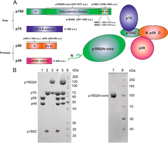
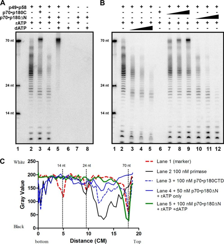
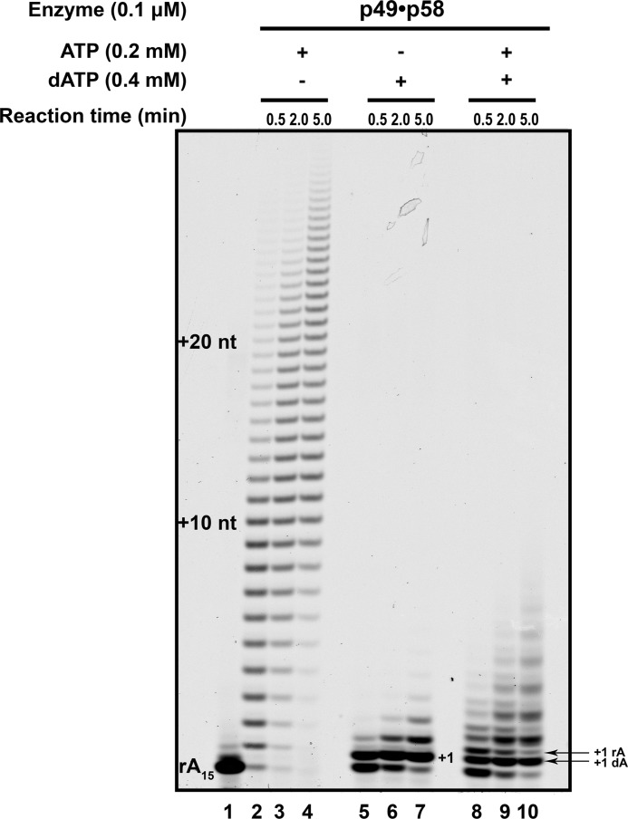
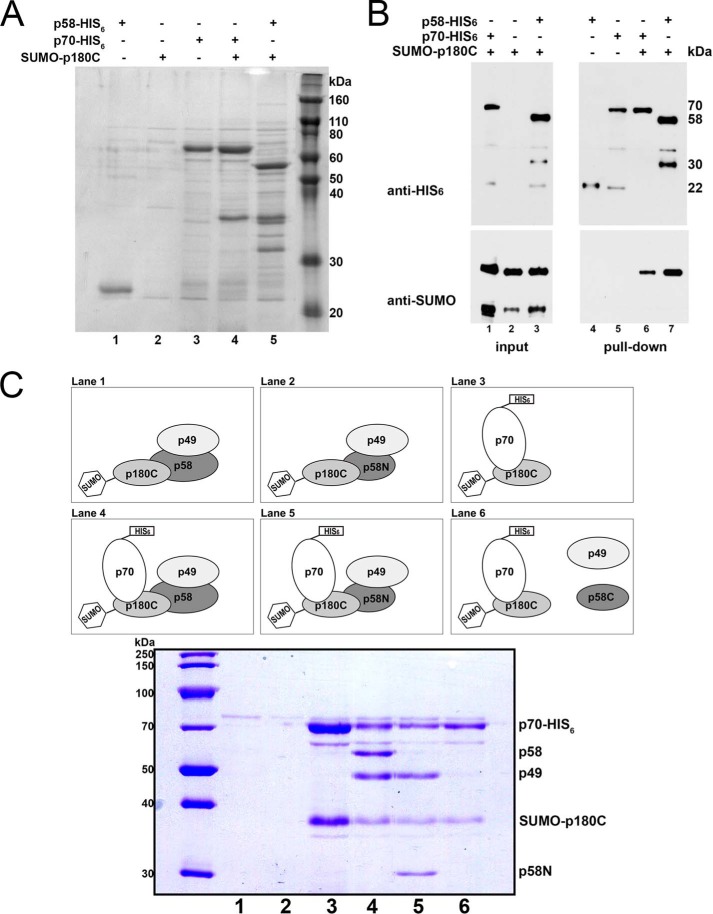
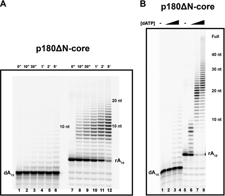
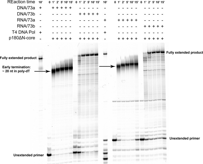
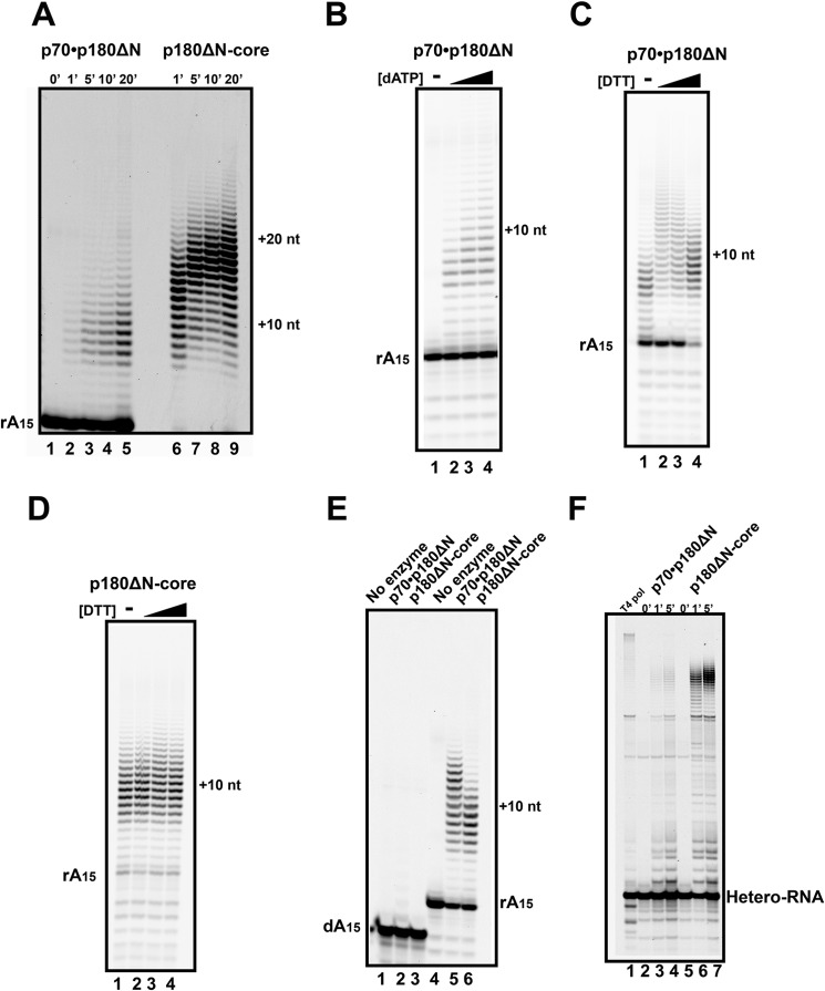
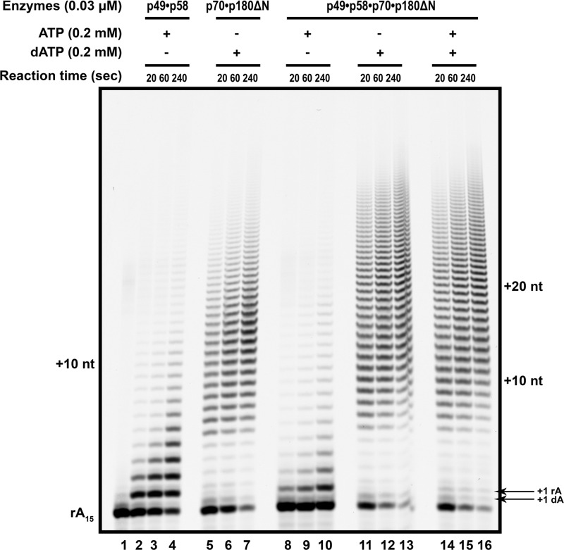
The initiation of DNA synthesis during replication of the human genome is accomplished primarily by the DNA polymerase α-primase complex, which makes the RNA-DNA primers accessible to processive DNA pols. The structural information needed to understand the mechanism of regulation of this complex biochemical reaction is incomplete. The presence of two enzymes in one complex poses the question of how these two enzymes cooperate during priming of DNA synthesis. Yeast two-hybrid and direct pulldown assays revealed that the N-terminal domain of the large subunit of primase (p58N) directly interacts with the C-terminal domain of the catalytic subunit of polα (p180C). We found that a complex of the C-terminal domain of the catalytic subunit of polα with the second subunit (p180C-p70) stimulated primase activity, whereas the whole catalytically active heterodimer of polα (p180ΔN-p70) inhibited RNA synthesis by primase. Conversely, the polα catalytic domain without the C-terminal part (p180ΔN-core) possessed a much higher propensity to extend the RNA primer than the two-subunit polα (p180ΔN-p70), suggesting that p180C and/or p70 are involved in the negative regulation of DNA pol activity. We conclude that the interaction between p180C, p70, and p58 regulates the proper primase and polymerase function. The composition of the template DNA is another important factor determining the activity of the complex. We have found that polα activity strongly depends on the sequence of the template and that homopyrimidine runs create a strong barrier for DNA synthesis by polα.

Keywords: C-terminal Domain; DNA Polymerase; DNA Polymerase Activity; DNA Polymerase α-Primase; DNA Primase; DNA Replication; DNA Replication Initiation; Primase Activity; Protein-Protein Interaction; RNA.


Figures

FIGURE 1.
FIGURE 1.
Overall structure of human polα-primase. A, schematic representation of the four-subunit complex, p180 (green), p70 (blue), p58 (orange), and p49 (red). The primary amino acids sequence and the domains of each subunit and various recombinant constructs studied in the current work are shown as stick diagrams on the left. Metal binding motifs in p180 are marked by yellow vertical lines and named two metal binding sites MBS1 and MBS2. B, analysis of the purity of human primase and polα samples. Left panel: lane 1, p49·p58; lane 2, p70·p180C; lane 3, p70·p180ΔN; lane 4, p49·p58·p70·p180C; lane 5, p49·p58·p70·p180ΔN; lane 6, EZ-Run Rec protein ladder (Fisher). Samples were run on 10% SDS-PAGE, and proteins were detected by Coomassie Blue staining. The p180ΔN corresponds to the catalytic subunit with deleted 291 amino acids from the N terminus. Right panel: lane 1, p180ΔN-core, lacking CTD of the catalytic subunit; lane 2, Page Ruler protein ladder (Thermo Scientific). Samples were run on 8% SDS-PAGE and visualized by Coomassie Blue staining.
FIGURE 2.
FIGURE 2.
Regulation of de novo primase activity by polα. polα-primase activity assay was done on a poly-dT 70-mer template (0.1 μm) as described under “Experimental Procedures.” A, effect of polα on primase activity. Lane 1, nucleotide size marker; lane 2, RNA primers synthesized by 0.1 μm p49·p58 alone; lane 3, RNA primers synthesized by 0.1 μm p49·p58 with the addition of 0.1 μm p70·p180C; lane 4, RNA primers synthesized by 0.1 μm p49·p58 with the addition of 0.05 μm p70·p180ΔN; lane 5, hybrid RNA-DNA fragments synthesized by 0.1 μm p49·p58·p70·p180ΔN complex with both the rATP and the dATP in reaction; lanes 6–8, negative controls. nt, nucleotides. B, primase activity assays with the titration of the polα variants. Lane 1, nucleotide size marker; lane 2, RNA primers synthesized by p49·p58 alone; lanes 3–5, RNA synthesized by p49·p58 in the presence of increasing concentrations of dATP (25, 50, 100 μm); lane 6, no activity by p70·p180C with rATP (negative control); lanes 7–9, the effect of increasing concentrations of p70·p180C (0.025–0.1 μm) on RNA synthesized by p49·p58; lane 10-12, RNA synthesized by p49·p58 with 0.05–0.2 μm p70·p180ΔN. C, quantification of gel shown in panel A. The mean gray value of each lane was selected from the bottom to the top of the gel image and analyzed by Image J. The distribution of the band (x axis: distance from bottom to the top of the gel) versus the intensity of the band (y axis: gray value closer to 0 means black, 250 means white).
FIGURE 3.
FIGURE 3.
Primase robustly extends RNA primers only in the presence of ribonucleotides. The poly-rA15 primer (enzyme to primer/template ratio = 1:1.5) was extended by heterodimeric primase (p49·p58) in the presence of rATP, dATP, or both. Lane 1, reaction without nucleotides; lanes 2–4, reaction with 0.2 mm ATP for 0.5, 2.0, and 5.0 min, respectively; lanes 5–7, reaction with 0.4 mm dATP for 0.5, 2.0, and 5.0 min, respectively; lanes 8–10, reaction with 0.2 mm ATP plus 0.4 mm dATP for 0.5, 2.0, and 5.0 min, respectively. nt, nucleotides.
FIGURE 4.
FIGURE 4.
N terminus of the large subunit of primase tethers it to polα. Analysis of the pulldown of proteins separated on SDS-PAGE and stained by Coomassie Blue (R250) (see “Experimental Procedures”). A, direct interaction between p58 and p180C. Lane 1, p58-His6 is self-cleaved to a 24-kDa product in the absence of its binding partner; lane 2, SUMO-p180C does not bind to Ni-IDA resins; lane 3, p70-His6 pulldown by Ni-IDA resin; lane 4, SUMO-p180C binds to p70-His6; lane 5, p180C sumo binds to p58-His6. B, Western blot detection of the interacting proteins seen in A. C, finding a region of p58 that is responsible for its interaction with polα. Lanes 1 and 2, p49, p58, p58N, or SUMO-p180C do not bind to Ni-IDA resin in the absence of p70-His6 (negative controls); lane 3, SUMO-p180C binds to p70-His6 (positive control). Lane 4, p58·p49 interacts with polα (SUMO-p180C·p70-His6; positive control); lane 5, p49·p58N interacts with SUMO-p180C·p70-His6; lane 6 shows the absence of interaction of p58C or p49 with SUMO-p180C·p70-His6.
FIGURE 5.
FIGURE 5.
Preferential RNA primer extension by human polα polymerase domain (p180ΔN-core) on the homopolymeric (dT)70 template. A, extension of the poly-dA15 and poly-rA15 primers (enzyme:primer/template ratios = 1:50 and 1:80, respectively) in the presence of 0.2 mm dATP. The products of reactions at the indicated time points were analyzed as described under “Experimental Procedures.” B, the dependence of the extension of the poly-dA15 and poly-rA15 primers (enzyme:primer/template ratios = 1:50 and 1:80, respectively) on the dATP concentration. The black triangle indicates that the reactions were carried out with the increase of dATP concentrations (0.01, 0.1, and 1 mm, respectively). nt, nucleotides. The reaction time was 20 min.
FIGURE 6.
FIGURE 6.
Same efficiency of the extension of DNA and RNA primers on hetero-homopolymeric hybrid and heteropolymeric DNA templates by the p180ΔN-core. For control of the full extension of the primers, we used reactions with T4 DNA polymerase, which robustly and completely extended DNA as well as RNA primers (left lane in the left and right halves of the gel). Arrows show the zones of termination of DNA synthesis. All reactions contain 0.2 mm dNTPs, and the enzyme to primer/template ratio was 1:50, except for the T4 DNA pol (∼1:2500).
FIGURE 7.
FIGURE 7.
Inhibitory effect of CTD and p70 on the primer extension by polα. A, the time course of the extension of the poly-rA15 primers on the poly-dT70 template by p70·p180ΔN (enzyme to primer/template ratio = 1:50; lane 2-5) or by the p180ΔN-core (enzyme to primer/template ratio = 1:100; lanes 6–9). nt, nucleotides. B, extension of the poly-rA15 primers by p70·p180ΔN on the poly-dT70 template (enzyme to primer/template ratio = 1:50, reaction time was 5 min). The black triangle indicates that the reactions were carried out with the increase of dATP concentrations of 0.01, 0.1, and 1 mm, respectively. C and D, effect of DTT on the ability of polα to extend the poly-rA15 primer. C, polα p70·p180ΔN (enzyme to primer/template ratio = 1:50, reaction time 10 in). D, p180ΔN-core (enzyme to primer/template ratio = 1:100, reaction time 5 min). The black triangle indicates that the reactions were carried out with the increase of DTT concentrations of 0, 0.5, 1.0, 2.0 mm DTT. E, smaller fragments in primer extension by excess of dimeric polα (p70·p180ΔN) in comparison to the polα core. Extension of the poly-dA15 and poly-rA15 primers in long reactions by the excess of p70·p180ΔN (enzyme to primer/template ratio = 1:50, reaction time 15 min) or by the p180ΔN-core (enzyme to primer/template ratio = 1:250, reaction time 2 min) with 0.2 mm dATP. F, lower activity of dimeric polα in comparison to the polα core on hybrid homo-heteropolymeric DNA template. Extension of the RNA primers on the DNA template 73a by p70·p180ΔN (0.025 μm) or by p180ΔN-core (enzyme to primer/template ratio = 1:50) for the indicated time points is shown. As a control, T4 DNA polymerase (∼1:2500, reaction time 2 min) was used to obtain a completely extended, full-length product.
FIGURE 8.
FIGURE 8.
Primase and DNA polymerase activities of the tetrameric polα-primase complex during the extension of the RNA primer on the polydT template. The poly-rA15 primer annealed to polydT and extended by primase (lanes 2–4: 0.2 mm rATP), polα (lanes 5–7: 0.2 mm dATP), or the polα-prim complex (lanes 8–16) is shown. Lane 1, reaction without nucleotides; lanes 8–10, reactions containing 0.2 mm rATP; lanes 11–13, reactions containing 0.2 mm dATP; lanes 14–16, reactions containing 0.2 mm mixture of rATP and dATP. All reactions have an enzyme to primer/template ratio of 1:15 for balanced RNA and DNA pols activities. In each case, the time course of the reaction included points of 20, 60, and 240 s. nt, nucleotides.

Similar articles

Cited by

References

    1. García-Gómez S., Reyes A., Martínez-Jiménez M. I., Chocrón E. S., Mourón S., Terrados G., Powell C., Salido E., Méndez J., Holt I. J., Blanco L. (2013) PrimPol, an archaic primase/polymerase operating in human cells. Mol. Cell 52, 541–553 - PMC - PubMed
    1. Garg P., Burgers P. M. (2005) DNA polymerases that propagate the eukaryotic DNA replication fork. Crit. Rev. Biochem. Mol. Biol. 40, 115–128 - PubMed
    1. Pavlov Y. I., Shcherbakova P. V., Rogozin I. B. (2006) Roles of DNA polymerases in replication, repair, and recombination in eukaryotes. Int. Rev. Cytol. 255, 41–132 - PubMed
    1. Kunkel T. A. (2009) Evolving views of DNA replication (in)fidelity. Cold Spring Harb. Symp. Quant. Biol. 74, 91–101 - PMC - PubMed
    1. MacNeill S. (2012) Composition and dynamics of the eukaryotic replisome: a brief overview. Subcell. Biochem. 62, 1–17 - PubMed

Publication types

MeSH terms

LinkOut - more resources