. 2014 Aug 29;289(35):24521-32.
doi: 10.1074/jbc.M114.579326. Epub 2014 Jun 24.

Substrate specificity of cytoplasmic N-glycosyltransferase

Affiliations

Substrate specificity of cytoplasmic N-glycosyltransferase

Andreas Naegeli et al. J Biol Chem. .

Abstract

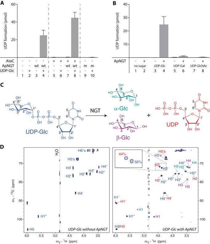
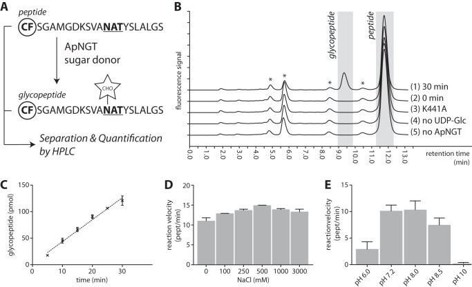
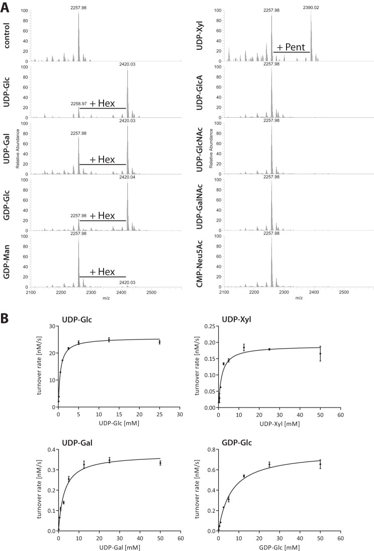
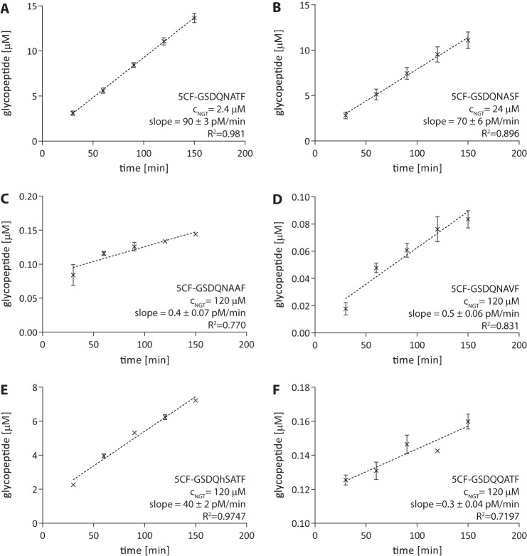
N-Linked protein glycosylation is a very common post-translational modification that can be found in all kingdoms of life. The classical, highly conserved pathway entails the assembly of a lipid-linked oligosaccharide and its transfer to an asparagine residue in the sequon NX(S/T) of a secreted protein by the integral membrane protein oligosaccharyltransferase. A few species in the class of γ-proteobacteria encode a cytoplasmic N-glycosylation system mediated by a soluble N-glycosyltransferase (NGT). This enzyme uses nucleotide-activated sugars to modify asparagine residues with single monosaccharides. As these enzymes are not related to oligosaccharyltransferase, NGTs constitute a novel class of N-glycosylation catalyzing enzymes. To characterize the NGT-catalyzed reaction, we developed a sensitive and quantitative in vitro assay based on HPLC separation and quantification of fluorescently labeled substrate peptides. With this assay we were able to directly quantify glycopeptide formation by Actinobacillus pleuropneumoniae NGT and determine its substrate specificities: NGT turns over a number of different sugar donor substrates and allows for activation by both UDP and GDP. Quantitative analysis of peptide substrate turnover demonstrated a strikingly similar specificity as the classical, oligosaccharyltransferase-catalyzed N-glycosylation, with NX(S/T) sequons being the optimal NGT substrates.

Keywords: Actinobacillus Pleuropneumoniae; Bacteria; Glycosylation; Glycosyltransferase; Post-translational Modification (PTM); Substrate Specificity.


Figures

FIGURE 1.
FIGURE 1.
ApNGT hydrolyzes nucleotide-activated sugars. A, substrate protein AtaC1866–2428 was incubated with UDP-Glc and purified ApNGT for 3 h at 30 °C and free UDP was determined using the bioluminescent UDP-Glo glycosyltransferase assay. Note that UDP is also formed in the absence of substrate protein but not in the absence of ApNGT or UDP-Glc or by the catalytically inactive ApNGT-K441A mutant. B, hydrolysis of different UDP-activated sugars. ApNGT hydrolyzed UDP-Glc and UDP-Gal (∼25 times slower) but not UDP-GlcNAc. C, schematic representation of UDP-Glc hydrolysis reaction. The color code corresponds to the color label in the NMR spectra. Numbers indicate the nomenclature of the protons. D, 1H-13C heteronuclear single-quantum coherence spectrum of 1 mm UDP-Glc measured with 2 scans at 293 K (left) and 1H-13C heteronuclear single-quantum coherence spectrum of 1 mm UDP-Glc after incubation for 20 h with 10 μm NGT measured with 32 scans at 293 K (right). The labels of the different reaction products are color coded corresponding to panel C. The assigned chemical shifts of UDP-Glc and UDP match previously reported values (45).
FIGURE 2.
FIGURE 2.
A direct assay for NGT activity. A, schematic overview of assay set up. A fluorescently labeled peptide (CF, 5-carboxyfluorescein) is incubated with purified ApNGT and a sugar donor substrate. Product (glycopeptide) is separated from educt (peptide) using RP-HPLC, and both peptides are quantified separately. B, representative chromatogram of analysis of glycosylation of peptide COK_139457–76. The peptide was incubated for 30 min with UDP-Glc and purified ApNGT. Samples were analyzed before (2) and after incubation time (1). The following controls were included: peptide with UDP-Glc and ApNGT-K441A (3), peptide with ApNGT alone (4), and peptide with UDP-Glc alone (5). The unmodified peptide elutes after 11.75 min, glycosylation decreases the retention time to 9.3 min. Glycosylation was further confirmed by MALDI mass spectrometry. Contaminants are marked with an asterisks. C, determination of the reaction speed of glycosylation. D and E, determination of the reaction speeds of NGT reaction at different salt concentrations and different pH values.
FIGURE 3.
FIGURE 3.
ApNGT is able to utilize different nucleotide-activated sugar donor substrates. A, 50 μm peptide COK_139457–76 was incubated with 5 μm purified ApNGT and 2 mm of the different nucleotide-activated sugars and the reaction products were analyzed by MALDI mass spectrometry. The unmodified peptide was detected at [M + H]+ = 2257.98. Upon incubation with UDP-Glc, UDP-Gal, GDP-Glc, and GDP-Man (left panel), an additional peak corresponding to the hexosylated peptide ([M + H]+ = 2420.03) was detected. Incubation with UDP-Xyl resulted in a peak corresponding to peptide modified with a pentose ([M + H]+ = 2390.02). No modification of the peptide with GlcA ([M + H]+ = 2434.01), GlcNAc or GalNAc ([M + H]+ = 2461.06), or Neu5Ac ([M + H]+ = 2549.08) could be detected. B, determination of Michaelis-Menten kinetics of turnover of different sugar donor substrates (UDP-Glc, UDP-Gal, UDP-Xyl, and GDP-Glc). Data were fitted using non-linear regression according to the Michaelis-Menten formula (R2 = 0.9884, 0.9637, 0.9345, and 0.9769, respectively). The kinetic parameters are summarized in Table 1.
FIGURE 4.
FIGURE 4.
Quantification of turnover rates of different substrate peptides. Determination of turnover rates of glycosylation reaction with different synthetic peptides. The amount of glycopeptide at each time point was determined based on HPLC separation and fluorescence detection. Each measurement was performed in triplicate. Parameters derived from this analysis are listed in Table 2.
FIGURE 5.
FIGURE 5.
Basic amino acids around the active site of APNGT are not involved in catalysis. A, crystal structure of ApNGT in complex with UDP (PBD code 3Q3H) (22). All basic residues in an 8-Å radius from the β-phosphate of the UDP moiety (in yellow) are marked in magenta. B, structure-aided sequence alignments of several GT41 glycosyltransferase sequences. The region around the residues marked in A is shown. For full alignment see supplemental Fig. S2. Alignment was performed using Expresso (28). Coloring indicates conservation of residues (from blue, not conserved, to red, fully conserved). C, in vivo activity assay of ApNGT mutants. The different mutants of ApNGT were co-expressed in E. coli with acceptor substrate AtaC1866–2428 and whole cell protein extracts were analyzed by immunoblot. NGT proteins were detected via the Myc epitope (top panel), AtaC1866–2428 via the His10 tag (middle panel), and glycosylation was detected using Asn-Glc specific serum MS14 (bottom panel) (23, 32).
FIGURE 6.
FIGURE 6.
In vitro analysis of ApNGT mutants H227A and K441A. 50 μm peptide COK_139457–76 was incubated with 10 mm UDP-Glc and 0.04 μm purified ApNGT (or the indicated mutant). Samples were taken every 10 min and analyzed by RP-HPLC. Turnover rates were determined by analyzing the data using linear regression and are depicted in brackets below the legend. For the control without any NGT enzyme and ApNGT-K441A no activity could be detected (n.d.)

Similar articles

Cited by

References

    1. Apweiler R., Hermjakob H., Sharon N. (1999) On the frequency of protein glycosylation, as deduced from analysis of the SWISS-PROT database. Biochim. Biophys. Acta 1473, 4–8 - PubMed
    1. Aebi M., Bernasconi R., Clerc S., Molinari M. (2010) N-Glycan structures: recognition and processing in the ER. Trends Biochem. Sci. 35, 74–82 - PubMed
    1. Helenius A., Aebi M. (2004) Roles of N-linked glycans in the endoplasmic reticulum. Annu. Rev. Biochem. 73, 1019–1049 - PubMed
    1. Sharon N., Lis H. (1995) Lectins–proteins with a sweet tooth: functions in cell recognition. Essays Biochem. 30, 59–75 - PubMed
    1. Kaneko Y., Nimmerjahn F., Ravetch J. V. (2006) Anti-inflammatory activity of immunoglobulin G resulting from Fc sialylation. Science 313, 670–673 - PubMed

Publication types

Substances