

Use of contrast-enhanced fluid-attenuated inversion recovery sequence to detect brain lesions in dogs and cats
- PMID: 24962604
- PMCID: PMC4857937
- DOI: 10.1111/jvim.12384
Use of contrast-enhanced fluid-attenuated inversion recovery sequence to detect brain lesions in dogs and cats
Abstract
Background: The diagnostic value of a contrast-enhanced T2-weighted FLAIR sequence (ceFLAIR) in brain imaging is unclear.
Hypothesis/objectives: That the number of brain lesions detected with ceFLAIR would be no greater than the sum of lesions detected with nFLAIR and ceT1W sequence.
Animals: One hundred and twenty-nine animals (108 dogs and 21 cats) undergoing magnetic resonance imaging (MRI) of the head between July 2010 and October 2011 were included in the study.
Methods: A transverse ceFLAIR was added to a standard brain MRI protocol. Presence and number of lesions were determined based on all available MRI sequences by 3 examiners in consensus and lesion visibility was evaluated for nFLAIR, ceFLAIR, and ceT1W sequences.
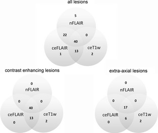
Results: Eighty-three lesions (58 intra-axial and 25 extra-axial) were identified in 51 patients. Five lesions were detected with nFLAIR alone, 2 with ceT1W alone, and 1 with ceFLAIR alone. Significantly higher numbers of lesions were detected using ceFLAIR than nFLAIR (76 versus 67 lesions; P = 0.04), in particular for lesions also detected with ceT1W images (53 versus 40; P =.01). There was no significant difference between the number of lesions detected with combined nFLAIR and ceT1W sequences compared to those detected with ceFLAIR (82 versus 76; P =.25).
Conclusion and clinical importance: Use of ceFLAIR as a complementary sequence to nFLAIR and ceT1W sequences did not improve the detection of brain lesions and cannot be recommended as part of a routine brain MRI protocol in dogs and cats with suspected brain lesions.
Keywords: Contrast enhancement; Intracranial lesions; Magnetic resonance imaging.
Copyright © 2014 by the American College of Veterinary Internal Medicine.
Figures
Similar articles
-
Exclusion of a brain lesion: is intravenous contrast administration required after normal precontrast magnetic resonance imaging?J Vet Intern Med. 2014 Mar-Apr;28(2):522-8. doi: 10.1111/jvim.12300. Epub 2014 Jan 27. J Vet Intern Med. 2014. PMID: 24467361 Free PMC article.
-
Contrast-enhanced fluid-attenuated inversion recovery vs. contrast-enhanced spin echo T1-weighted brain imaging.Vet Radiol Ultrasound. 2008 Jul-Aug;49(4):333-8. doi: 10.1111/j.1740-8261.2008.00375.x. Vet Radiol Ultrasound. 2008. PMID: 18720762
-
Use of the T2*-weighted gradient recalled echo sequence for magnetic resonance imaging of the canine and feline brain.Vet Radiol Ultrasound. 2014 Nov-Dec;55(6):599-606. doi: 10.1111/vru.12164. Epub 2014 May 16. Vet Radiol Ultrasound. 2014. PMID: 24833062
-
MRI of brain disease in veterinary patients part 2: Acquired brain disorders.Vet Clin North Am Small Anim Pract. 2010 Jan;40(1):39-63. doi: 10.1016/j.cvsm.2009.09.006. Vet Clin North Am Small Anim Pract. 2010. PMID: 19942056 Review.
-
Quantitative MRI for brain lesion diagnosis in dogs and cats: A comprehensive overview.Vet Radiol Ultrasound. 2024 Nov;65(6):849-864. doi: 10.1111/vru.13434. Epub 2024 Sep 27. Vet Radiol Ultrasound. 2024. PMID: 39329277 Review.
Cited by
-
Abrogation of fluid suppression in intracranial postcontrast fluid-attenuated inversion recovery magnetic resonance imaging: A clinical and phantom study.Vet Radiol Ultrasound. 2018 Jul;59(4):432-443. doi: 10.1111/vru.12605. Epub 2018 Feb 8. Vet Radiol Ultrasound. 2018. PMID: 29424062 Free PMC article.
References
-
- Benigni L, Lamb CR. Comparison of fluid‐attenuated inversion recovery and T2‐weighted magnetic resonance images in dogs and cats with suspected brain disease. Vet Radiol Ultrasound 2005;46:287–292. - PubMed
-
- Brant‐Zawadzki M, Atkinson D, Detrick M, et al. Fluid‐attenuated inversion recovery (FLAIR) for assessment of cerebral infarction. Initial clinical experience in 50 patients. Stroke 1996;27:1187–1191. - PubMed
-
- Demaerel P, Bosmans H, Caerts B, et al. Fast FLAIR MRI in childhood white‐matter abnormalities. Neuroradiology 1998;40:355–358. - PubMed
-
- Falzone C, Rossi F, Calistri M, et al. Contrast‐enhanced fluid‐attenuated inversion recovery vs. contrast‐enhanced spin echo T1‐weighted brain imaging. Vet Radiol Ultrasound 2008;49:333–338. - PubMed
MeSH terms
Substances
LinkOut - more resources
Full Text Sources
Other Literature Sources
Medical
Miscellaneous
