. 2014 Aug;93(4):682-97.
doi: 10.1111/mmi.12681. Epub 2014 Jul 16.

CarD integrates three functional modules to promote efficient transcription, antibiotic tolerance, and pathogenesis in mycobacteria

Affiliations

CarD integrates three functional modules to promote efficient transcription, antibiotic tolerance, and pathogenesis in mycobacteria

Ashley L Garner et al. Mol Microbiol. 2014 Aug.

Abstract

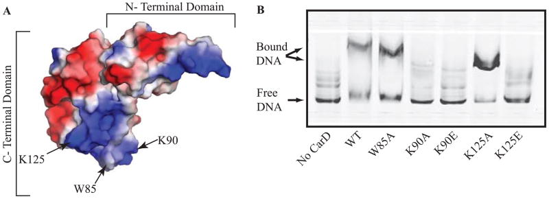
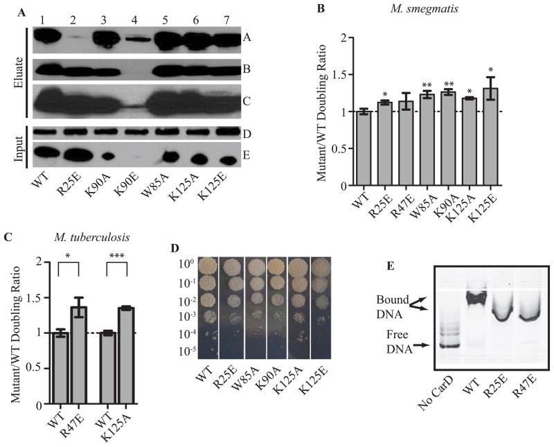
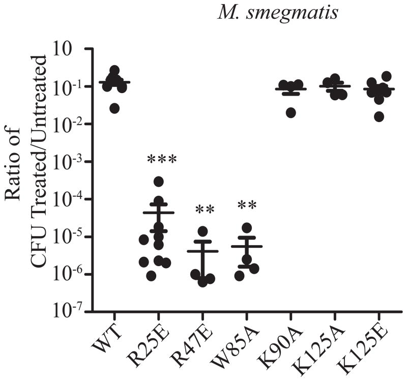
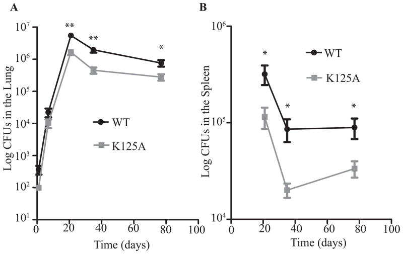
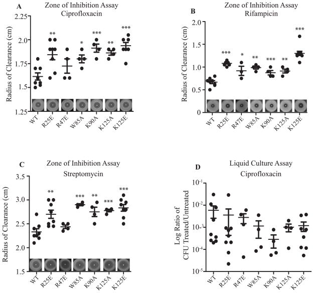
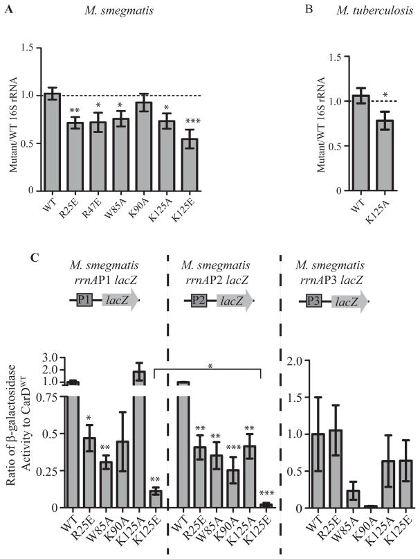
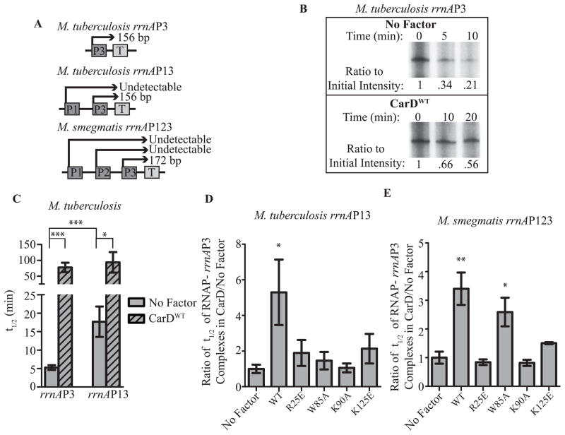
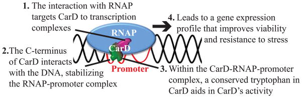
Although the basic mechanisms of prokaryotic transcription are conserved, it has become evident that some bacteria require additional factors to allow for efficient gene transcription. CarD is an RNA polymerase (RNAP)-binding protein conserved in numerous bacterial species and essential in mycobacteria. Despite the importance of CarD, its function at transcription complexes remains unclear. We have generated a panel of mutations that individually target three independent functional modules of CarD: the RNAP interaction domain, the DNA-binding domain, and a conserved tryptophan residue. We have dissected the roles of each functional module in CarD activity and built a model where each module contributes to stabilizing RNAP-promoter complexes. Our work highlights the requirement of all three modules of CarD in the obligate pathogen Mycobacterium tuberculosis, but not in Mycobacterium smegmatis. We also report divergent use of the CarD functional modules in resisting oxidative stress and pigmentation. These studies provide new information regarding the functional domains involved in transcriptional regulation by CarD while also improving understanding of the physiology of M. tuberculosis.


Conflict of interest statement

Conflict of Interest

The authors declare no conflict of interest.

Figures

Figure 1
Figure 1. CarD’s C-terminal basic patch is responsible for its interaction with DNA
A. Electrostatic surface diagram of M. tuberculosis CarD modified from (Srivastava et al., 2013) illustrating the location of W85, K90, and K125 within the basic patch. B. Image of a nondenaturing polyacrylamide gel from an EMSA with no protein, M. smegmatis CarDWT, CarDW85A, CarDK90A, CarDK90E, CarDK125A, or CarDK125E incubated with IRDye labeled M. smegmatis rrnAPL DNA. The reactions were separated on a nondenaturing polyacrylamide gel, which was then imaged using the Odyssey CLX imaging system (LI-COR).
Figure 2
Figure 2. Each of CarD’s functional domains is required for optimal growth in mycobacteria
A. Immunoprecipitation experiments with a monoclonal antibody specific for HA in the M. smegmatis strains expressing CarDWT-HA (lane 1), CarDR25E-HA (lane 2), CarDK90A-HA (lane 3), CarDK90E-HA (lane 4), CarDW85A-HA (lane 5), CarDK125A-HA (lane 6), or CarDK125E-HA (lane 7). Inputs (before immunoprecipitation) and eluates were analyzed by western blotting with antibodies specific for either RNAP β (Panel A and D) or CarD (Panel B, C, and E). Panel C is a longer exposure of the film from panel B in order to show the CarDK90E band. B–C. The doubling time of each CarD strain was expressed as a ratio to the doubling time of the CarDWT expressing strain for the M. smegmatis strains expressing CarDWT, CarDR25E, CarDK90A, CarDW85A, CarDK125A, or CarDK125E (B) and M. tuberculosis strains expressing CarDWT, CarDK125A, or CarDR47E (C). Each graph shows the mean ± SEM of data from at least three replicates. Significance of the differences between mutant strains and WT were determined by calculating P values by Student’s t test. An asterisk indicates significance with a P value of <0.05, two asterisks indicate significance with a P value of <0.01, and three asterisks indicate significance with a P value of <0.005. D. Plated dilutions of M. smegmatis strains expressing CarDWT, CarDR25E, CarDW85A, CarDK90A, CarDK125A, or CarDK125E on LB after 3 days of growth at 37°C. E. Image of nondenaturing polyacrylamide gel from EMSAs with no protein, M. tuberculosis CarDWT, CarDR25E, or CarDR47E incubated with IRDye labeled M. smegmatis rrnAPL DNA. The reactions were separated on a nondenaturing polyacrylamide gel, which was then imaged using the Odyssey CLX imaging system (LI-COR).
Figure 3
Figure 3
The CarD-DNA interaction is dispensable for resistance to killing by oxidative stress. Log-phase M. smegmatis strains expressing CarDWT, CarDR25E, CarDR47E, CarDW85A, CarDK90A, CarDK125A, or CarDK125E in LB were treated for 1 hr with 25 mM H2O2. After treatment, dilutions were plated on LB and the surviving CFUs were counted. Survival of each replicate is graphed as a ratio of CFU in treated cultures to that in untreated cultures along with the mean ± SEM of the ratios for each set of replicates. Each sample is represented by a black circle.
Figure 4
Figure 4
A mutation in the DNA binding domain of CarD affects the pathogenesis of M. tuberculosis in murine tissues. C57BL/6 mice were infected by the aerosol route with the M. tuberculosis strains expressing either CarDWT (black circles) or CarDK125A (grey squares). Shown are bacterial titers in the lungs (A) and spleens (B) of the infected mice. Each time point is the mean ± SEM of data from 6 mice per strain, combined from two experiments. The significance of the differences between the CarDK125A and the CarDWT strain in both panels were determined as described for Figure 2.
Figure 5
Figure 5. Each of CarD’s functional domains is important for resistance to clinically relevant antibiotics
A–C. Survival of M. smegmatis strains during the disk zone of inhibition assays. Five hundred microliters of log-phase M. smegmatis strains expressing CarDWT, CarDR25E, CarDR47E, CarDW85A, CarDK90A, CarDK125A, or CarDK125E were plated on LB agar, and a disk spotted with 5μl of 1 mg ml−1 ciprofloxacin (A), 100 mg ml−1 rifampicin (B), or 200 mg ml−1 streptomycin (C) was placed in the middle of the bacterial lawn. A representative experiment is shown above the x-axis. The radius of the zone of inhibition for each replicate and the mean ± SEM for each set of replicates is graphed, with each sample represented by a black circle. D. Survival of M. smegmatis strains during transient ciprofloxacin treatment. Log-phase cultures of the M. smegmatis strains expressing CarDWT, CarDR25E, CarDR47E, CarDW85A, CarDK90A, CarDK125A, or CarDK125E growing in LB were treated for 2 hr with 10 μg ml−1 ciprofloxacin before the dilutions were plated to determine the surviving CFU. Graphed is the ratio of CFUs in treated cultures to that in untreated cultures for each replicate and the mean ± SEM for each set of replicates, with each sample represented by a black circle. For all panels, the significance of differences between mutant strains and WT were determined as described in Figure 2.
Figure 6
Figure 6. Each of CarD’s functional domains are important for transcriptional regulation
A–B. 16S rRNA levels in log phase cultures of M. smegmatis strains expressing CarDWT, CarDR25E, CarDR47E, CarDW85A, CarDK90A, CarDK125A, or CarDK125E grown in LB (A) and M. tuberculosis strains expressing CarDWT or CarDK125A grown in 7H9 (B) as determined by qRT-PCR and expressed as a ratio to the levels in CarDWT expressing strains. Each graph shows the mean ± SEM of data from at least 3 replicates and the significance of the differences between mutant strains and WT. C. M. smegmatis strains expressing CarDWT, CarDR25E, CarDR47E, CarDW85A, CarDK90A, CarDK125A, or CarDK125E were transformed with the promoter-lacZ fusion constructs diagramed above each graph. The ratio of β-galactosidase activity that resulted from lacZ expression from the M. smegmatis rrnAP1, rrnAP2, or rrnAP3 promoter in each fusion in the CarD mutant versus CarDWT expressing strains was calculated and graphed as the mean of at least 3 replicates ± SEM. Shown above each bar is the significance of the difference between the mutant strain and WT from the same promoter. The bracket designates the significance of the difference between the activation of the rrnAP1 and rrnAP2 promoters in the CarDK125E expressing strain. For all panels, the significance of differences were calculated as described in Figure 2.
Figure 7
Figure 7. CarD’s interaction with DNA is important for stabilization of RNAP-promoter complexes
A. Schematic of the M. tuberculosis and M. smegmatis rRNA promoter constructs used during in vitro transcription assays. The length of the rrnAP3-derived transcripts initiated at the promoter and ending at the terminator (T) are shown. B. Autoradiographs of 32P-labeled transcripts on denaturing polyacrylamide gels from a representative complex stability assay using M. bovis RNAP-σA holoenzyme and the rrnAP3 construct. The amount of transcript formed at the indicated timepoints following the addition of competitor is shown when no factor is added versus when CarDWT is added. The intensity of each band was quantified and the ratio to the intensity at time 0 was calculated and annotated under the gel. C. The half-life (t1/2) of RNAP-promoter complexes formed at the rrnAP3 promoter in the M. tuberculosis rrnAP3 and the M. tuberculosis rrnAP13 constructs in the presence (hatched bars) and absence (solid grey bars) of CarDWT was calculated in GraphPad Prism based on the amount of transcripts formed during complex stability assays. The graph shows the mean of at least 3 replicates ± SEM and the significance of the differences in the comparisons designated by the brackets are shown. D–E. Ratio of the t1/2 of RNAP-promoter complexes containing CarDWT, CarDR25E, CarDW85A, CarDK90A, or CarDK125E to the t1/2 of RNAP-promoter complexes in the absence of CarD as determined by single round complex stability in vitro transcription assays at the rrnAP3 promoter on the M. tuberculosis rrnAP13 construct (D) or the M. smegmatis rrnAP123 construct (E). The significance of differences as compared to the reactions containing no factor are shown. For all panels, the significance of differences were determined as described for Figure 2.
Figure 8
Figure 8
Model of CarD activity at promoters. CarD is targeted to the initiation complex via its interaction with RNAP-β (1). The C-terminus of CarD then interacts with the DNA (2). The combined association of CarD with the RNAP and CarD with the promoter DNA stabilizes the RNAP-promoter complex. Within the CarD-RNAP-promoter complex, the conserved tryptophan in CarD performs a function to facilitate CarD activity (3). The efficient functioning of all three of CarD’s activities improves viability and resistance to stress and results in changes in gene expression (4).

Similar articles

Cited by

References

    1. Bremer H, Dennis PP. Escherichia Coli and Salmonella Typhimurium: Cellular and Molecular Biology. ASM Press; 1996. Modulation of Chemical Composition and Other Parameters of the Cell by Growth Rate; pp. 1553–69.
    1. Chao MC, Rubin EJ. Letting sleeping dos lie: does dormancy play a role in tuberculosis? Annu Rev Microbiol. 2010;64:293–311. - PubMed
    1. Cooper AM, Segal BH, Frank AA, Holland SM, Orme IM, Orme IANM. Transient Loss of Resistance to Pulmonary Tuberculosis in p47 phox −/− Mice Transient Loss of Resistance to Pulmonary Tuberculosis. Infect Immun. 2000;68:1231–1234. - PMC - PubMed
    1. Czyz A, Mooney RA, Iaconi A. Mycobacterial RNA Polymerase Requires a U-Tract at Intrinsic Terminators and Is Aided by NusG at Suboptimal Terminators. MBio. 2014;5:1–10. - PMC - PubMed
    1. Gaal T, Ross W, Estrem ST, Nguyen LH, Burgess RR, Gourse RL. Promoter recognition and discrimination by EsigmaS RNA polymerase. Mol Microbiol. 2001;42:939–54. - PubMed

Publication types

MeSH terms

LinkOut - more resources